

Volume 10, Issue 2 (5-2023)
JROS 2023, 10(2): 109-114 |
Back to browse issues page


Download citation:
BibTeX | RIS | EndNote | Medlars | ProCite | Reference Manager | RefWorks
Send citation to:


BibTeX | RIS | EndNote | Medlars | ProCite | Reference Manager | RefWorks
Send citation to:
Kargar Shooroki K, Mokhles P, Toloue Ghamari B, Komijani M. A Rare Case of Intraosseous Spindle Cell Hemangioendothelioma in the Ischium. JROS 2023; 10 (2) :109-114
URL: http://jros.iums.ac.ir/article-1-2237-en.html
URL: http://jros.iums.ac.ir/article-1-2237-en.html


1- Department of Orthopedics, Bone and Joint Reconstruction Research Center, School of Medicine, Shafayahyaeian Hospital, Iran University of Medical Science, Tehran, Iran.
2- Department of Pathology, School of Medicine, Iran University of Medical Science, Tehran, Iran.
3- Department of Orthopedics, School of Medicine, Iran University of Medical Sciences, Tehran, Iran.
2- Department of Pathology, School of Medicine, Iran University of Medical Science, Tehran, Iran.
3- Department of Orthopedics, School of Medicine, Iran University of Medical Sciences, Tehran, Iran.
Full-Text [PDF 1606 kb]
(215 Downloads)
| Abstract (HTML) (954 Views)
Full-Text: (183 Views)
Introduction
Spindle cell hemangioendothelioma (SCH) is a rare benign tumor characterized by thin-walled, cavernous, and vascular spaces. Some spaces contain phleboliths, while others are filled with cellular stroma composed of spindled fibroblastic cells. Weiss and Enzinger were the first to mention this in 1986 [1]. Although initially considered a moderately malignant vascular tumor due to its tendency for local recurrence, SCH’s malignant potential was later re-evaluated and found to have a low metastatic potential. This type of tumor is most commonly found in soft tissues, but its occurrence in bone tissues is infrequent [2]. Typically, spindle cell hemangiomas are superficial or subcutaneous erythematous nodules that tend to appear in the limbs of young individuals, particularly the hands, with an equal sex distribution [3]. SCH pathologic features include cavernous blood vessels and solid regions of spindle and epithelioid cells [4]. It was formerly assumed to be a tumor with limited metastatic potential, but it is now classified as a benign vascular tumor rather than a low-grade angiosarcoma [5].
This article aims to introduce a known case of intraosseous SCH lesion in a 55-year-old woman in the ischium bone who presented with pelvic pain.
Case Presentation
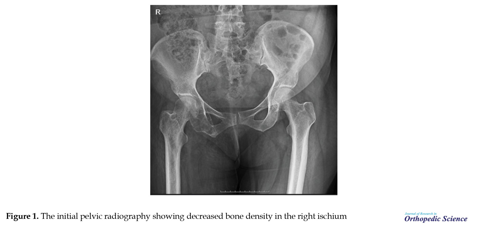
A 55-year-old woman presented with pelvic pain that was mostly felt on the right side. Pelvic radiography showed a significant decrease in the density of the right ischium (Figure 1).
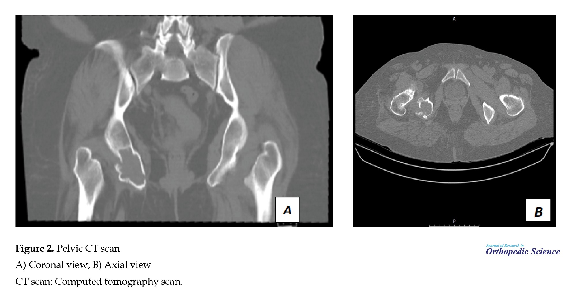
 Computed tomography (CT) scan of the ischium indicated a lytic expanding lesion in the ischium (Figure 2).
 Pathology showed a spindle cell area after the patient underwent a CT-guided biopsy.
Surgical procedure
After the pathological results were determined, a bone curettage surgical plan was arranged for the patient. Following spinal anesthesia, the patient was placed in a prone position with the hips and knees lightly flexed. An incision was made through the gluteus maximus and hamstring muscles down to the ischium. Curettage was performed, and a bone graft was placed.
Pathology
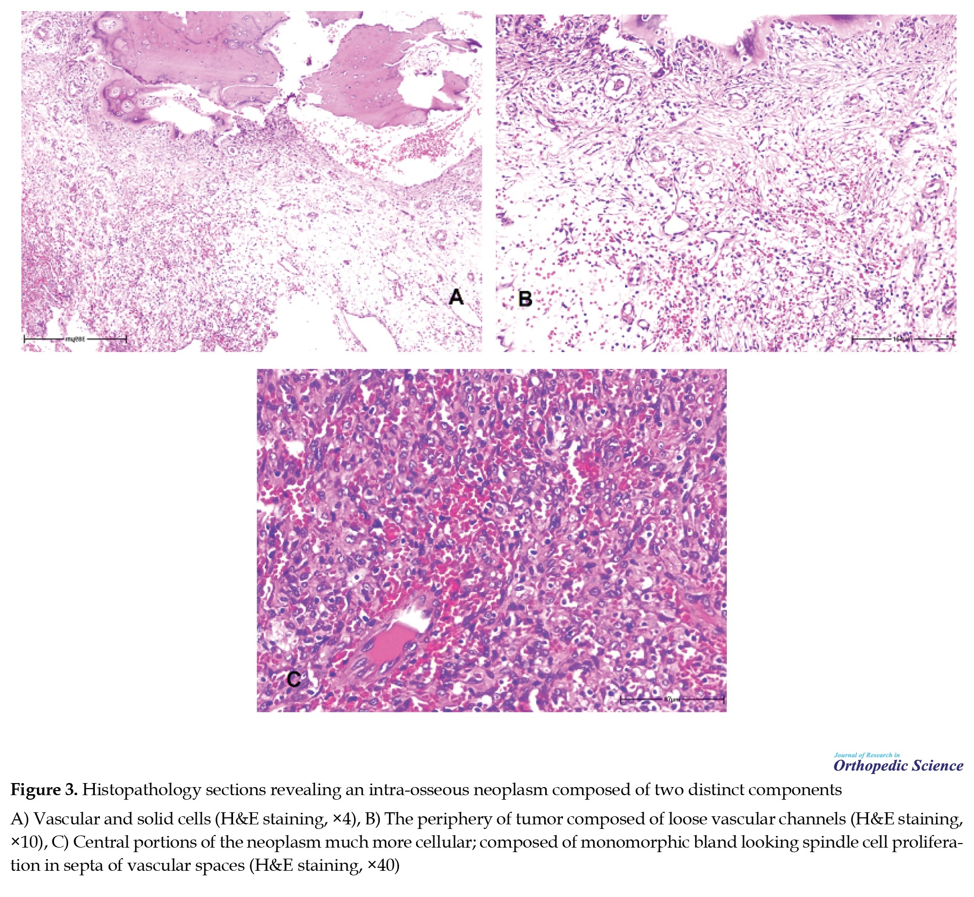
A sample was extracted for histological study, and the results showed an intra-osseous neoplasm composed of two distinct components: Vascular and solid cells. The periphery of the tumor exhibited loose vascular channels intermingled with a centrally located hypercellular area, consisting of monomorphic, bland-looking spindle cell proliferation in septa of vascular spaces (Figure 3).
Follow-up
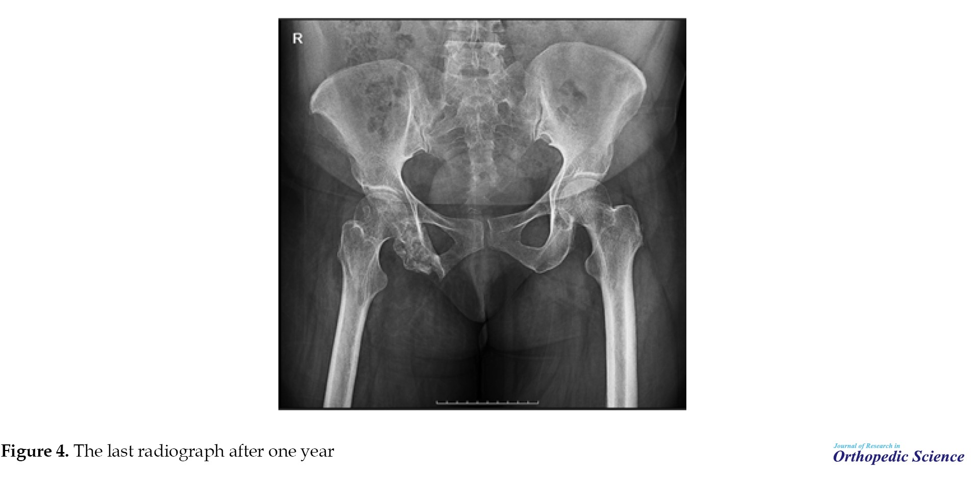
The patient was followed-up for one year and had no complaints. The last pelvic radiography was performed one year after surgery, and complete healing of the bone tissue was evident (Figure 4).
Discussion
Tumors originating from bone vasculature are rare, constituting less than 1% of all bone tumors [6]. There are three types of tumors currently classified as epithelioid endothelial cell tumors - epithelioid hemangioma, epithelioid hemangioendothelioma, and epithelioid angiosarcoma [7]. A SCH may also contain a small component of epithelioid endothelial cells. Weiss and Enzinger first described a new type of vascular tumor called “spindle cell hemangioendothelioma” in 1986. This type of tumor has limited malignant potential and is now known as SCH, a benign and often multifocal neoplasm. It typically affects young adults and develops in the subcutaneous tissue of the distal extremities, particularly the hands. In contrast to other hemangiomas, SCH rarely develops in the bone [8]. SCH is histologically characterized as epithelioid and spindle cell hemangioma by the presence of dilated capillaries bordered with flattened endothelial cells filled with red cells, similar to those found in cavernous hemangiomas. These capillaries were tightly mixed with spindle cell components. Furthermore, in SCH, epithelioid cells align in solid cords rather than lining well-formed vascular gaps, similar to epithelioid and spindle cell hemangiomas [2, 9]. Only a few reports of intraosseous spindle cell hemangiomas or hemangioendotheliomas have been published in the medical literature. Spindle cell hemangiomas have been reported in the calcaneus [10], fibula periosteum [11], and frontal bones [12, 13]. Additionally, one case of SCH in the sacrum has been reported [14].
Conclusion
We now describe a rare case of SCH of the ischium. The radiologic differential diagnosis for this case includes several types of tumors, including giant cell tumors of the bone, aneurysmal bone cysts, metastatic bone tumors, solitary bone cysts, and vascular tumors, such as hemangiomas. The patient was managed by surgical curettage and bone grafting; a one-year follow-up showed promising results and was pain-free.
Ethical Considerations
Compliance with ethical guidelines
There were no ethical considerations to be considered in this research.
Funding
This research did not receive any grant from funding agencies in the public, commercial, or non-profit sectors.
Authors' contributions
All authors contributed equally to the conception and design of the study, data collection and analysis, interception of the results and drafting of the manuscript. Each author approved the final version of the manuscript for submission.
Conflict of interest
The authors declared no conflict of interest.
References
Spindle cell hemangioendothelioma (SCH) is a rare benign tumor characterized by thin-walled, cavernous, and vascular spaces. Some spaces contain phleboliths, while others are filled with cellular stroma composed of spindled fibroblastic cells. Weiss and Enzinger were the first to mention this in 1986 [1]. Although initially considered a moderately malignant vascular tumor due to its tendency for local recurrence, SCH’s malignant potential was later re-evaluated and found to have a low metastatic potential. This type of tumor is most commonly found in soft tissues, but its occurrence in bone tissues is infrequent [2]. Typically, spindle cell hemangiomas are superficial or subcutaneous erythematous nodules that tend to appear in the limbs of young individuals, particularly the hands, with an equal sex distribution [3]. SCH pathologic features include cavernous blood vessels and solid regions of spindle and epithelioid cells [4]. It was formerly assumed to be a tumor with limited metastatic potential, but it is now classified as a benign vascular tumor rather than a low-grade angiosarcoma [5].
This article aims to introduce a known case of intraosseous SCH lesion in a 55-year-old woman in the ischium bone who presented with pelvic pain.
Case Presentation
A 55-year-old woman presented with pelvic pain that was mostly felt on the right side. Pelvic radiography showed a significant decrease in the density of the right ischium (Figure 1).
Surgical procedure
After the pathological results were determined, a bone curettage surgical plan was arranged for the patient. Following spinal anesthesia, the patient was placed in a prone position with the hips and knees lightly flexed. An incision was made through the gluteus maximus and hamstring muscles down to the ischium. Curettage was performed, and a bone graft was placed.
Pathology
A sample was extracted for histological study, and the results showed an intra-osseous neoplasm composed of two distinct components: Vascular and solid cells. The periphery of the tumor exhibited loose vascular channels intermingled with a centrally located hypercellular area, consisting of monomorphic, bland-looking spindle cell proliferation in septa of vascular spaces (Figure 3).
Follow-up
The patient was followed-up for one year and had no complaints. The last pelvic radiography was performed one year after surgery, and complete healing of the bone tissue was evident (Figure 4).
Discussion
Tumors originating from bone vasculature are rare, constituting less than 1% of all bone tumors [6]. There are three types of tumors currently classified as epithelioid endothelial cell tumors - epithelioid hemangioma, epithelioid hemangioendothelioma, and epithelioid angiosarcoma [7]. A SCH may also contain a small component of epithelioid endothelial cells. Weiss and Enzinger first described a new type of vascular tumor called “spindle cell hemangioendothelioma” in 1986. This type of tumor has limited malignant potential and is now known as SCH, a benign and often multifocal neoplasm. It typically affects young adults and develops in the subcutaneous tissue of the distal extremities, particularly the hands. In contrast to other hemangiomas, SCH rarely develops in the bone [8]. SCH is histologically characterized as epithelioid and spindle cell hemangioma by the presence of dilated capillaries bordered with flattened endothelial cells filled with red cells, similar to those found in cavernous hemangiomas. These capillaries were tightly mixed with spindle cell components. Furthermore, in SCH, epithelioid cells align in solid cords rather than lining well-formed vascular gaps, similar to epithelioid and spindle cell hemangiomas [2, 9]. Only a few reports of intraosseous spindle cell hemangiomas or hemangioendotheliomas have been published in the medical literature. Spindle cell hemangiomas have been reported in the calcaneus [10], fibula periosteum [11], and frontal bones [12, 13]. Additionally, one case of SCH in the sacrum has been reported [14].
Conclusion
We now describe a rare case of SCH of the ischium. The radiologic differential diagnosis for this case includes several types of tumors, including giant cell tumors of the bone, aneurysmal bone cysts, metastatic bone tumors, solitary bone cysts, and vascular tumors, such as hemangiomas. The patient was managed by surgical curettage and bone grafting; a one-year follow-up showed promising results and was pain-free.
Ethical Considerations
Compliance with ethical guidelines
There were no ethical considerations to be considered in this research.
Funding
This research did not receive any grant from funding agencies in the public, commercial, or non-profit sectors.
Authors' contributions
All authors contributed equally to the conception and design of the study, data collection and analysis, interception of the results and drafting of the manuscript. Each author approved the final version of the manuscript for submission.
Conflict of interest
The authors declared no conflict of interest.
References
- Weiss SW, Enzinger FM. Spindle cell hemangioendothelioma. A low-grade angiosarcoma resembling a cavernous hemangioma and Kaposi's sarcoma. Am J Surg Pathol. 1986; 10(8):521-30. [DOI:10.1097/00000478-198608000-00001] [PMID]
- Perkins P, Weiss SW. Spindle cell hemangioendothelioma. An analysis of 78 cases with reassessment of its pathogenesis and biologic behavior. Am J Surg Pathol. 1996; 20(10):1196-204. [DOI:10.1097/00000478-199610000-00004] [PMID]
- Weiss SW, Goldblum JR, Folpe AL. Enzinger and Weiss’s soft tissue tumors. Edinburgh: Elsevier Health Sciences; 2007. [Link]
- Weiss SW, Goldblum JR. Spindle cell hemangioma. In: Weiss SW, & Goldblum JR, editors. Enzinger and Weiss’s soft tissue tumors. Philadelphia: Mosby/Elsevier; 2008. [Link]
- Ding J, Hashimoto H, Imayama S, Tsuneyoshi M, Enjoji M. Spindle cell haemangioendothelioma: probably a benign vascular lesion not a low-grade angiosarcoma. A clinicopathological, ultrastructural and immunohistochemical study. Virchows Arch A Pathol Anat Histopathol. 1992; 420(1):77-85. [DOI:10.1007/BF01605988] [PMID]
- Boutin RD, Spaeth HJ, Mangalik A, Sell JJ. Epithelioid hemangioendothelioma of bone. Skeletal Radiol. 1996; 25(4):391-5. [DOI:10.1007/s002560050102] [PMID]
- Schajowicz F. Histologic typing of bone tumors. Geneva: World Health Organization; 1993. [DOI:10.1007/978-3-642-84902-2]
- Maclean FM, Schatz J, McCarthy SW, Scolyer RA, Stalley P, Bonar SF. Epithelioid and spindle cell haemangioma of bone. Skeletal Radiol. 2007; 36(Suppl 1):S50-7. [DOI:10.1007/s00256-006-0135-z] [PMID]
- Keel SB, Rosenberg AE. Hemorrhagic epithelioid and spindle cell hemangioma: A newly recognized, unique vascular tumor of bone. Cancer. 1999; 85(9):1966-72. [DOI: 10.1002/(SICI)1097-0142(19990501)85:9<1966::AID-CNCR13>3.0.CO;2-W]
- Hakozaki M, Tajino T, Watanabe K, Yamada H, Kikuchi S, Hojo H, et al. Intraosseous spindle cell hemangioma of the calcaneus: A case report and review of the literature. Ann Diagn Pathol. 2012; 16(5):369-73. [DOI:10.1016/j.anndiagpath.2012.01.005] [PMID]
- Tsukamoto S, Honoki K, Shimada K, Fujii H, Kido A, Takano M, et al. Periosteal spindle cell hemangioma of the fibula: A case report. Skeletal Radiol. 2013; 42(8):1165-8. [DOI:10.1007/s00256-013-1603-x] [PMID]
- Jun Wu, You Zhou, Jun Fan. Skull spindle cell hemangioma: Case report. Chin J Neurosurg Dis Res. 2011; 10:280-1.
- Huang C, Zhang H, Guan L, Luo J. Rare spindle cell hemangioma of bone: Case report and literature review. Radiol Case Rep. 2022; 17(3):886-890. [DOI:10.1016/j.radcr.2021.11.051] [PMID] [PMCID]
- Winter A, Siu A, Jamshidi A, Malawer M, Sherman JH. Spindle cell hemangioendothelioma of the sacrum: Case report. J Neurosurg Spine. 2014; 21(2):275-8. [DOI:10.3171/2014.3.SPINE13651] [PMID]
Type of Study: Case Report |
Subject:
Tumor surgery
Received: 2022/04/19 | Accepted: 2022/05/27 | Published: 2023/05/1
Received: 2022/04/19 | Accepted: 2022/05/27 | Published: 2023/05/1
| Rights and permissions | |
 |
This work is licensed under a Creative Commons Attribution-NonCommercial 4.0 International License. |

Copyright © The Author(s);
This is an open access article distributed under the terms of the Creative Commons Attribution License (CC-By-NC), which permits use, distribution, and reproduction in any medium, provided the original work is properly cited and is not used for commercial purposes.
Contact Information
