

Volume 13, Issue 1 (Winter 2026)
JROS 2026, 13(1): 25-30 |
Back to browse issues page




Download citation:
BibTeX | RIS | EndNote | Medlars | ProCite | Reference Manager | RefWorks
Send citation to:


BibTeX | RIS | EndNote | Medlars | ProCite | Reference Manager | RefWorks
Send citation to:
Khajeh Alizadeh Attar M, Nakhaei Amroodi M, Bahaeddini M, Mahdavifar M, Tabrizian P. Anatomical Neck Fracture of the Humerus: Two Rare Cases and Functional Outcomes. JROS 2026; 13 (1) :25-30
URL: http://jros.iums.ac.ir/article-1-2315-en.html
URL: http://jros.iums.ac.ir/article-1-2315-en.html
Mojtaba Khajeh Alizadeh Attar1 
 , Morteza Nakhaei Amroodi1 , Mohammadreza Bahaeddini1 , Mohammadmehdi Mahdavifar1 , Pouria Tabrizian1
, Morteza Nakhaei Amroodi1 , Mohammadreza Bahaeddini1 , Mohammadmehdi Mahdavifar1 , Pouria Tabrizian1
1- Bone and Joint Reconstruction Research Center, School of Medicine, Shafayahyaeian Hospital, Iran University of Medical Sciences, Tehran, Iran.
Keywords: Proximal humerus fracture, Anatomical neck fracture, Lesser tuberosity, Internal fixation, Shoulder injury
Full-Text [PDF 3887 kb]
(34 Downloads)
| Abstract (HTML) (481 Views)
Full-Text: (2 Views)
Introduction
Proximal humerus fractures account for approximately 4–5% of all fractures and are particularly common in the elderly; however, high-energy trauma in younger individuals can result in more complex injury patterns [1]. Among these, fractures involving the anatomical neck of the humerus are relatively rare but clinically significant due to their strong association with disruption of the humeral head blood supply and the consequent risk of avascular necrosis (AVN) [2, 3].
The vascularity of the humeral head is primarily supplied by the anterior and posterior circumflex humeral arteries, with the arcuate artery playing a key intraosseous role. Disruption of this delicate vascular network, particularly in anatomical neck fractures, significantly increases the risk of ischemia and subsequent osteonecrosis [3, 4]. Therefore, early diagnosis and appropriate management are critical for preserving joint function and preventing long-term complications.
Fractures of the lesser tuberosity are uncommon and are often associated with posterior shoulder dislocations or high-energy trauma. These concomitant injuries are rare and may be overlooked on initial radiographs, particularly if minimally displaced, and are better visualized using advanced imaging modalities such as computed tomography (CT) [5, 6]. The lesser tuberosity serves as the insertion site of the subscapularis tendon; thus, its involvement can lead to significant functional impairment, particularly affecting internal rotation and anterior shoulder stability [7].
The management of these rare fracture patterns remains controversial and depends on multiple factors, including patient age, bone quality, and surgeon’s experience. Surgical intervention is often indicated in displaced or complex fractures to restore anatomy, optimize functional outcomes, and reduce the probability of humeral head AVN [8, 9]. Open reduction and internal fixation (ORIF), commonly performed via the deltopectoral approach, remains a widely accepted technique, allowing for direct visualization and stable fixation of fracture fragments [10, 11].
Given the rarity of combined anatomical neck and lesser tuberosity fractures, limited data are available regarding optimal treatment strategies and outcomes. This case report presents the successful surgical management of a complex injury, highlighting the importance of accurate diagnosis, appropriate fixation techniques, and structured postoperative rehabilitation for achieving favorable clinical outcomes.
Case Presentation
Case 1: A 37-year-old right-hand-dominant male presented to the emergency department following a fall from a height of approximately 4 meters, resulting in trauma to his left shoulder. On initial examination, the patient exhibited marked swelling, severe limitation of shoulder motion, and localized tenderness. Initial radiographic evaluation was performed, followed by a CT scan for a more precise assessment (Figures 1A, 1B, 1C, 1D, 1E and 1F).
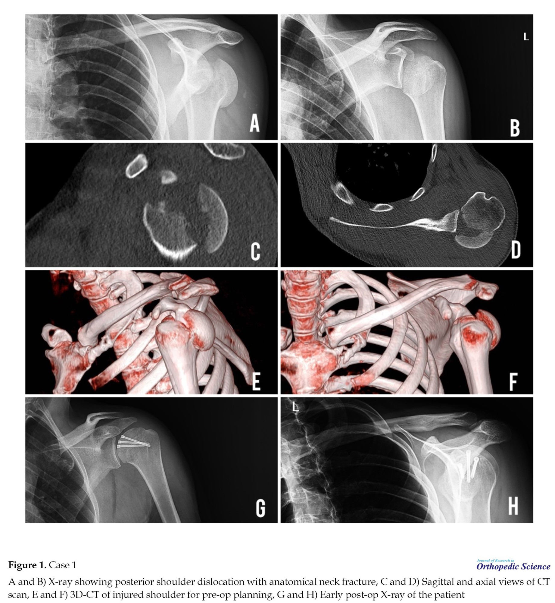 Imaging revealed a fracture of the anatomical neck of the humeral head associated with a fracture of the lesser tuberosity. Based on these findings, surgical management with internal fixation was indicated. The procedure was performed under general anesthesia using a deltopectoral approach. Fracture fragments were anatomically reduced. Fixation of the humeral head was achieved using Herbert screws, while the lesser tuberosity was secured using Ethibond sutures (Figure 1).
Case 2: A 24-year-old worker presented with right shoulder direct trauma due to a fall from the third floor. Initial examination showed an anatomical neck of humerus fracture and posterior dislocation which underwent ORIF. During surgery, approximately 80% of the humeral head cartilage surface was destroyed, and the head was reduced and fixed with Herbert screws (Figure 2).
 Postoperatively, passive shoulder movements and pendulum exercises were initiated two weeks after surgery. Gradual progression of range of motion was implemented through supervised physiotherapy and controlled rehabilitation protocols. Follow-up evaluations at 2 weeks, 4 weeks, 3 months, and 6 months demonstrated satisfactory fracture :union:, significant improvement in shoulder range of motion, and no radiological evidence of AVN. Clinical images demonstrating the patient’s range of motion have been provided (Figures 3A, 3B, 3C, and 3D).
 These cases highlight that accurate diagnosis, appropriate surgical technique, and structured rehabilitation can lead to favorable functional outcomes in complex fractures of the anatomical neck of the humerus.
Discussion
Fractures of the anatomical neck of the humerus are rare but clinically significant injuries due to their association with disruption of humeral head perfusion and the potential development of AVN [3]. Hertel et al. identified specific morphological predictors of ischemia, including disruption of the medial hinge and short calcar segment, highlighting that anatomical neck fractures are among the highest-risk patterns for humeral head necrosis [3].
Vascular compromise remains the most critical concern in these injuries. The blood supply of the humeral head is primarily derived from the anterior and posterior circumflex humeral arteries, with the arcuate artery contributing to intraosseous perfusion [4]. Gerber et al. demonstrated that injury patterns involving the anatomical neck may significantly disrupt this vascular network, predisposing to AVN if not managed appropriately [4]. However, several clinical studies have shown that early anatomical reduction and stable fixation can reduce the incidence of AVN even in high-risk fractures [12].
The associated fracture of the lesser tuberosity is an uncommon but functionally important injury. Ogawa and Takahashi reported that isolated lesser tuberosity fractures are frequently missed on plain radiographs and often require CT imaging for accurate diagnosis [6]. Because the subscapularis tendon inserts on the lesser tuberosity, its disruption can result in impaired internal rotation strength and anterior shoulder instability [6, 13]. In the present cases, fixation of the fragment allowed restoration of subscapularis function, contributing to favorable postoperative outcomes.
Surgical management of displaced proximal humerus fractures remains a topic of debate. The deltopectoral approach with ORIF is widely used because it provides excellent exposure for anatomical reduction and stable fixation [14]. Robinson et al. emphasized that restoration of anatomical alignment is the most important factor influencing functional outcomes in proximal humerus fractures, particularly in younger patients with high-energy trauma [14].
The timing of mobilization also plays a crucial role in functional recovery. Hertel et al. and other authors have shown that prolonged immobilization after proximal humerus fractures is associated with stiffness and poorer functional results, while early controlled mobilization improves shoulder function [3, 15]. In this case, early initiation of passive motion at two weeks likely contributed to the satisfactory restoration of range of motion.
Conclusion
Overall, these two cases support existing evidence that even complex proximal humerus fractures involving the anatomical neck and lesser tuberosity can achieve good outcomes if managed with early diagnosis, anatomical reduction, appropriate fixation, and structured rehabilitation.
Ethical Considerations
Compliance with ethical guidelines
Written informed consent was obtained from both two patients for using their medical information, radiographic images, and their photographs. The patient privacy was considered and their face in photograph was faded. The patients were free to leave the study whenever they wanted. This study was conducted in accordance with the ethical principles of the Declaration of Helsinki.
Funding
This research did not receive any grant from funding agencies in the public, commercial, or non-profit sectors.
Authors' contributions
Conceptualization and surgery: Pouria Tabrizian; Data collection: Mohammadmehdi Mahdavifar; Writing: Mojtaba Khajeh Alizadeh Attar and Mohammadreza Bahaeddini; Supervision: Morteza Nakhaei Amroodi.
Conflict of interest
The authors declared no conflict of interest.
References
Proximal humerus fractures account for approximately 4–5% of all fractures and are particularly common in the elderly; however, high-energy trauma in younger individuals can result in more complex injury patterns [1]. Among these, fractures involving the anatomical neck of the humerus are relatively rare but clinically significant due to their strong association with disruption of the humeral head blood supply and the consequent risk of avascular necrosis (AVN) [2, 3].
The vascularity of the humeral head is primarily supplied by the anterior and posterior circumflex humeral arteries, with the arcuate artery playing a key intraosseous role. Disruption of this delicate vascular network, particularly in anatomical neck fractures, significantly increases the risk of ischemia and subsequent osteonecrosis [3, 4]. Therefore, early diagnosis and appropriate management are critical for preserving joint function and preventing long-term complications.
Fractures of the lesser tuberosity are uncommon and are often associated with posterior shoulder dislocations or high-energy trauma. These concomitant injuries are rare and may be overlooked on initial radiographs, particularly if minimally displaced, and are better visualized using advanced imaging modalities such as computed tomography (CT) [5, 6]. The lesser tuberosity serves as the insertion site of the subscapularis tendon; thus, its involvement can lead to significant functional impairment, particularly affecting internal rotation and anterior shoulder stability [7].
The management of these rare fracture patterns remains controversial and depends on multiple factors, including patient age, bone quality, and surgeon’s experience. Surgical intervention is often indicated in displaced or complex fractures to restore anatomy, optimize functional outcomes, and reduce the probability of humeral head AVN [8, 9]. Open reduction and internal fixation (ORIF), commonly performed via the deltopectoral approach, remains a widely accepted technique, allowing for direct visualization and stable fixation of fracture fragments [10, 11].
Given the rarity of combined anatomical neck and lesser tuberosity fractures, limited data are available regarding optimal treatment strategies and outcomes. This case report presents the successful surgical management of a complex injury, highlighting the importance of accurate diagnosis, appropriate fixation techniques, and structured postoperative rehabilitation for achieving favorable clinical outcomes.
Case Presentation
Case 1: A 37-year-old right-hand-dominant male presented to the emergency department following a fall from a height of approximately 4 meters, resulting in trauma to his left shoulder. On initial examination, the patient exhibited marked swelling, severe limitation of shoulder motion, and localized tenderness. Initial radiographic evaluation was performed, followed by a CT scan for a more precise assessment (Figures 1A, 1B, 1C, 1D, 1E and 1F).
Case 2: A 24-year-old worker presented with right shoulder direct trauma due to a fall from the third floor. Initial examination showed an anatomical neck of humerus fracture and posterior dislocation which underwent ORIF. During surgery, approximately 80% of the humeral head cartilage surface was destroyed, and the head was reduced and fixed with Herbert screws (Figure 2).
Discussion
Fractures of the anatomical neck of the humerus are rare but clinically significant injuries due to their association with disruption of humeral head perfusion and the potential development of AVN [3]. Hertel et al. identified specific morphological predictors of ischemia, including disruption of the medial hinge and short calcar segment, highlighting that anatomical neck fractures are among the highest-risk patterns for humeral head necrosis [3].
Vascular compromise remains the most critical concern in these injuries. The blood supply of the humeral head is primarily derived from the anterior and posterior circumflex humeral arteries, with the arcuate artery contributing to intraosseous perfusion [4]. Gerber et al. demonstrated that injury patterns involving the anatomical neck may significantly disrupt this vascular network, predisposing to AVN if not managed appropriately [4]. However, several clinical studies have shown that early anatomical reduction and stable fixation can reduce the incidence of AVN even in high-risk fractures [12].
The associated fracture of the lesser tuberosity is an uncommon but functionally important injury. Ogawa and Takahashi reported that isolated lesser tuberosity fractures are frequently missed on plain radiographs and often require CT imaging for accurate diagnosis [6]. Because the subscapularis tendon inserts on the lesser tuberosity, its disruption can result in impaired internal rotation strength and anterior shoulder instability [6, 13]. In the present cases, fixation of the fragment allowed restoration of subscapularis function, contributing to favorable postoperative outcomes.
Surgical management of displaced proximal humerus fractures remains a topic of debate. The deltopectoral approach with ORIF is widely used because it provides excellent exposure for anatomical reduction and stable fixation [14]. Robinson et al. emphasized that restoration of anatomical alignment is the most important factor influencing functional outcomes in proximal humerus fractures, particularly in younger patients with high-energy trauma [14].
The timing of mobilization also plays a crucial role in functional recovery. Hertel et al. and other authors have shown that prolonged immobilization after proximal humerus fractures is associated with stiffness and poorer functional results, while early controlled mobilization improves shoulder function [3, 15]. In this case, early initiation of passive motion at two weeks likely contributed to the satisfactory restoration of range of motion.
Conclusion
Overall, these two cases support existing evidence that even complex proximal humerus fractures involving the anatomical neck and lesser tuberosity can achieve good outcomes if managed with early diagnosis, anatomical reduction, appropriate fixation, and structured rehabilitation.
Ethical Considerations
Compliance with ethical guidelines
Written informed consent was obtained from both two patients for using their medical information, radiographic images, and their photographs. The patient privacy was considered and their face in photograph was faded. The patients were free to leave the study whenever they wanted. This study was conducted in accordance with the ethical principles of the Declaration of Helsinki.
Funding
This research did not receive any grant from funding agencies in the public, commercial, or non-profit sectors.
Authors' contributions
Conceptualization and surgery: Pouria Tabrizian; Data collection: Mohammadmehdi Mahdavifar; Writing: Mojtaba Khajeh Alizadeh Attar and Mohammadreza Bahaeddini; Supervision: Morteza Nakhaei Amroodi.
Conflict of interest
The authors declared no conflict of interest.
References
- Court-Brown CM, Caesar B. Epidemiology of adult fractures: A review. Injury. 2006; 37(8):691-7. [DOI:10.1016/j.injury.2006.04.130] [PMID]
- Neer C. Displaced proximal humeral fractures: I. Classification and Evaluation. J Bone Joint Surg. 1970; 52:1077-89. [DOI:10.2106/00004623-197052060-00001]
- Hertel R, Hempfing A, Stiehler M, Leunig M. Predictors of humeral head ischemia after intracapsular fracture of the proximal humerus. J Shoulder Elbow Surg. 2004; 13(4):427-33. [DOI:10.1016/j.jse.2004.01.034] [PMID]
- Gerber C, Schneeberger A, Vinh T. The arterial vascularization of the humeral head. An anatomical study. J Bone Joint Surg. 1990; 72(10):1486-94. [DOI:10.2106/00004623-199072100-00009]
- Scheibel M, Martinek V, Imhoff AB. Arthroscopic reconstruction of an isolated avulsion fracture of the lesser tuberosity. Arthroscopy. 2005; 21(4):487-94. [DOI:10.1016/j.arthro.2004.11.012] [PMID]
- Ogawa K, Takahashi M. Long-term outcome of isolated lesser tuberosity fractures of the humerus. J Trauma. 1997; 42(5):955-9. [DOI:10.1097/00005373-199705000-00029] [PMID]
- Gerber C, Krushell RJ. Isolated rupture of the tendon of the subscapularis muscle. Clinical features in 16 cases. J Bone Joint Surg Br. 1991; 73(3):389-94. [DOI:10.1302/0301-620X.73B3.1670434] [PMID]
- Brorson S. Proximal humeral fractures. In: Court-Brown Ch, McQueen M, Swiontkowski MF, Ring D, Friedman SM, Duckworth AD, editors. Musculoskeletal trauma in the elderly. Boca Raton: CRC Press; 2016. [DOI:10.1201/9781315381954-20]
- Launonen AP, Lepola V, Saranko A, Flinkkilä T, Laitinen M, Mattila VM. Epidemiology of proximal humerus fractures. Arch Osteoporos. 2015; 10:209. [DOI:10.1007/s11657-015-0209-4] [PMID]
- Nho SJ, Brophy RH, Barker JU, Cornell CN, MacGillivray JD. Management of proximal humeral fractures based on current literature. J Bone Joint Surg Am. 2007; 89(Suppl 3):44-58. [DOI:10.2106/JBJS.G.00648] [PMID]
- Szyszkowrtz R, Seggl W, Schleifer P, Cundy PJ. Proximal humeral fractures: Management techniques and expected results. Clin Orthop Relat Res. 1993; 292:13-25. [DOI:10.1097/00003086-199307000-00005]
- Schnetzke M, Bockmeyer J, Loew M, Studier-Fischer S, Grützner PA, Guehring T. Rate of avascular necrosis after fracture dislocations of the proximal humerus: Timing of surgery. Obere Extrem. 2018; 13(4):273-8. [DOI:10.1007/s11678-018-0452-6]
- Lyons RP, Green A. Subscapularis tendon tears. J Am Acad Orthop Surg. 2005; 13(5):353-63. [DOI:10.5435/00124635-200509000-00009] [PMID]
- Robinson CM, Khan LA, Akhtar MA. Treatment of anterior fracture-dislocations of the proximal humerus by open reduction and internal fixation. J Bone Joint Surg Br. 2006; 88(4):502-8. [DOI:10.1302/0301-620X.88B4.17195] [PMID]
- Court-Brown CM, Garg A, McQueen MM. The epidemiology of proximal humeral fractures. Acta Orthop Scand. 2001; 72(4):365-71. [DOI:10.1080/000164701753542023] [PMID]
Type of Study: Case Report |
Subject:
Shoulder / Elbow
Received: 2025/12/11 | Accepted: 2026/01/14 | Published: 2026/01/1
Received: 2025/12/11 | Accepted: 2026/01/14 | Published: 2026/01/1
| Rights and permissions | |
 |
This work is licensed under a Creative Commons Attribution-NonCommercial 4.0 International License. |

Copyright © The Author(s);
This is an open access article distributed under the terms of the Creative Commons Attribution License (CC-By-NC), which permits use, distribution, and reproduction in any medium, provided the original work is properly cited and is not used for commercial purposes.
Contact Information
