

Volume 13, Issue 2 (Spring 2026)
JROS 2026, 13(2): 49-56 |
Back to browse issues page




Download citation:
BibTeX | RIS | EndNote | Medlars | ProCite | Reference Manager | RefWorks
Send citation to:


BibTeX | RIS | EndNote | Medlars | ProCite | Reference Manager | RefWorks
Send citation to:
Askari A, Jabalameli M, Moghtadaei M, Yahyazadeh H, Gholami E, Zareie B et al . Mid-term Outcomes of Lateral Opening-wedge Distal Femoral Osteotomy for Valgus Knees. JROS 2026; 13 (2) :49-56
URL: http://jros.iums.ac.ir/article-1-2322-en.html
URL: http://jros.iums.ac.ir/article-1-2322-en.html
Alireza Askari1 

 , Mahmoud Jabalameli1 , Mehdi Moghtadaei2 , Hooman Yahyazadeh3 , Ehsan Gholami1 , Bushra Zareie1 , Behnam Sour1
, Mahmoud Jabalameli1 , Mehdi Moghtadaei2 , Hooman Yahyazadeh3 , Ehsan Gholami1 , Bushra Zareie1 , Behnam Sour1
1- Department of Orthopedics, Bone and Joint Reconstruction Research Center, School of Medicine, Iran University of Medical Sciences, Tehran, Iran.
2- Department of Orthopedics, Rasoul Akram Hospital, Iran University of Medical Sciences, Tehran, Iran
3- Department of Orthopedic Surgery, Farhikhtegan Hospital, TeMS.C. Islamic Azad University, Tehran, Iran.
2- Department of Orthopedics, Rasoul Akram Hospital, Iran University of Medical Sciences, Tehran, Iran
3- Department of Orthopedic Surgery, Farhikhtegan Hospital, TeMS.C. Islamic Azad University, Tehran, Iran.
Keywords: Distal femoral osteotomy (DFO), Valgus knee, Lateral opening wedge, Alignment correction, Nonunion, 36-item short form health survey (SF-36)
Full-Text [PDF 2006 kb]
(47 Downloads)
| Abstract (HTML) (544 Views)
Full-Text: (16 Views)
Introduction
Valgus malalignment shifts the mechanical axis laterally and increases the contact pressure in the lateral compartment, contributing to pain and progressive degeneration of the lateral compartment. In young and active patients, arthroplasty may be less desirable due to activity restrictions and the risk of future revision. Distal femoral osteotomy (DFO) is a joint-preserving realignment procedure that aims to unload the lateral compartment by restoring a more neutral mechanical axis in knees with primarily femoral deformity [1–5].
DFO can be performed using a medial closing wedge or a lateral opening wedge technique. Systematic reviews suggest both approaches can achieve reliable correction and comparable outcomes when performed with careful preoperative planning and stable fixation; however, complication profiles differ, and delayed :union:/non:union: has been highlighted as a particular concern after lateral opening-wedge techniques [3–6]. Mid- to long-term series report that DFO can delay conversion to arthroplasty in appropriately selected patients, but survivorship and complication rates vary with age, baseline osteoarthritis severity, and treatment of the osteotomy gap [7, 8].
The purposes of this study were (1) to report radiographic correction, final pain and health-related quality of life, and complications after lateral opening-wedge DFO fixed with a locking compression plate (LCP); and (2) to assess factors associated with outcomes and complications. We hypothesized that DFO would restore near-neutral alignment with acceptable final visual analogue scale (VAS) and SF-36 scores, and that older age and higher K grade would be associated with worse outcomes and an increased risk of complications.
Methods
Study design and setting
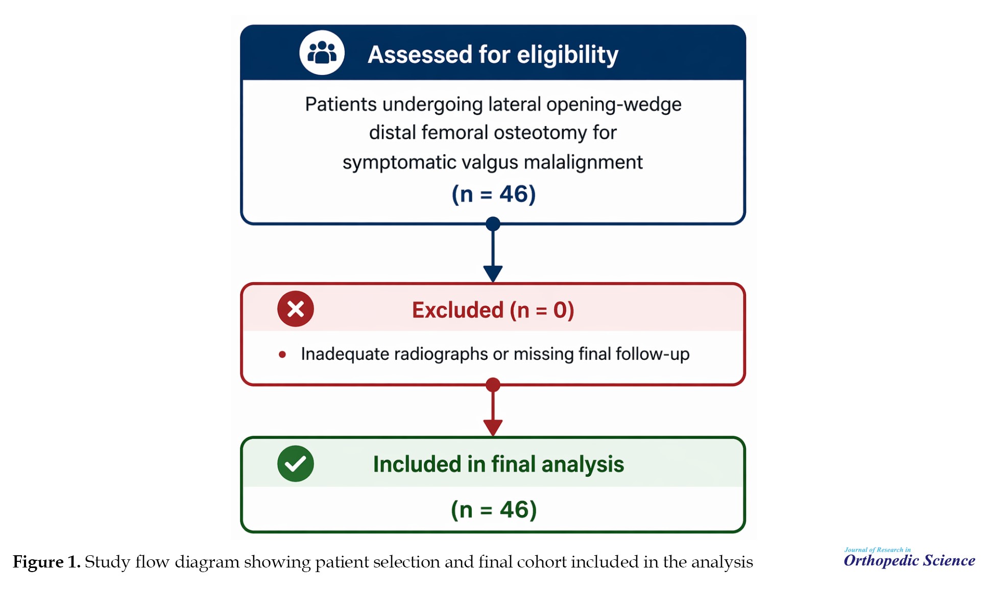
This retrospective cohort study was conducted at three tertiary referral hospitals. Data were extracted from institutional medical records between October 2016 and October 2024. The study was reported in accordance with the strengthening the reporting of observational studies in epidemiology (STROBE) statement for observational studies (Figure 1) [9].
Participants
Consecutive patients who underwent lateral opening-wedge DFO for symptomatic valgus malalignment were screened. The primary indication was symptomatic valgus alignment on standing long-leg radiographs (hip–knee–ankle [HKA]>180° as measured in our institution) with clinical and/or radiographic evidence of lateral compartment overload. Patients were included if a complete radiographic dataset (pre- and postoperative long-leg radiographs) and final follow-up clinical assessment were available. Patients with inflammatory arthropathy or advanced tricompartmental degeneration were not considered candidates for osteotomy [1, 2].
Surgical technique
All procedures were performed using a lateral opening-wedge DFO via a direct lateral approach, and the DFO was fixed with a lateral LCP. The goal was to restore near-neutral mechanical alignment based on preoperative planning and intraoperative fluoroscopic confirmation.
Postoperative rehabilitation
Range-of-motion exercises were started immediately after surgery as tolerated. Patients remained non-weight-bearing for approximately 6 weeks (1.5 months), followed by progressive loading. Full weight bearing was permitted after approximately 12 weeks (3 months), based on clinical assessment and radiographic review.
Radiographic assessment
Long-leg anteroposterior radiographs were used to evaluate lower-limb alignment. The HKA angle was measured preoperatively and at final follow-up. In this study, values greater than 180 degrees indicated valgus alignment [10]. Preoperative osteoarthritis severity was classified using the Kellgren–Lawrence (KL) grading system on weight-bearing radiographs [11].
Clinical outcomes
Pain at final follow-up was assessed using a VAS, 0–10 [12]. Health-related quality of life at final follow-up was assessed using the 36-item short form health survey (SF-36, 0–100) [13]. Preoperative VAS and SF-36 scores were unavailable in the medical records and were not analyzed.
Complications and failure
Postoperative complications were identified by chart review. In this cohort, complications comprised non:union: and conversion to total knee arthroplasty (TKA). Failure was defined a priori as conversion to arthroplasty (TKA) during follow-up [6].
Statistical analysis
Continuous variables are reported as Mean±SD and range, and categorical variables as counts and percentages. Pre- versus postoperative HKA was compared using a paired t-test. Correlations were assessed using Pearson correlation (age vs SF-36) and Spearman correlation (KL grade vs SF-36). Differences in VAS across KL grades were tested using one-way analysis of variance (ANOVA). Comparisons between patients with and without complications were performed using independent-samples t-tests for continuous variables and Fisher exact tests for categorical variables. Statistical significance was set at P<0.05. Analyses were performed using IBM SPSS software, version 16 (IBM Corp., Armonk, NY, USA).
Use of generative AI tools
Generative AI tools were used only for language refinement during manuscript preparation. All scientific content, analysis, and interpretations were developed and verified by the authors, who take full responsibility for the manuscript.
Results
Cohort characteristics
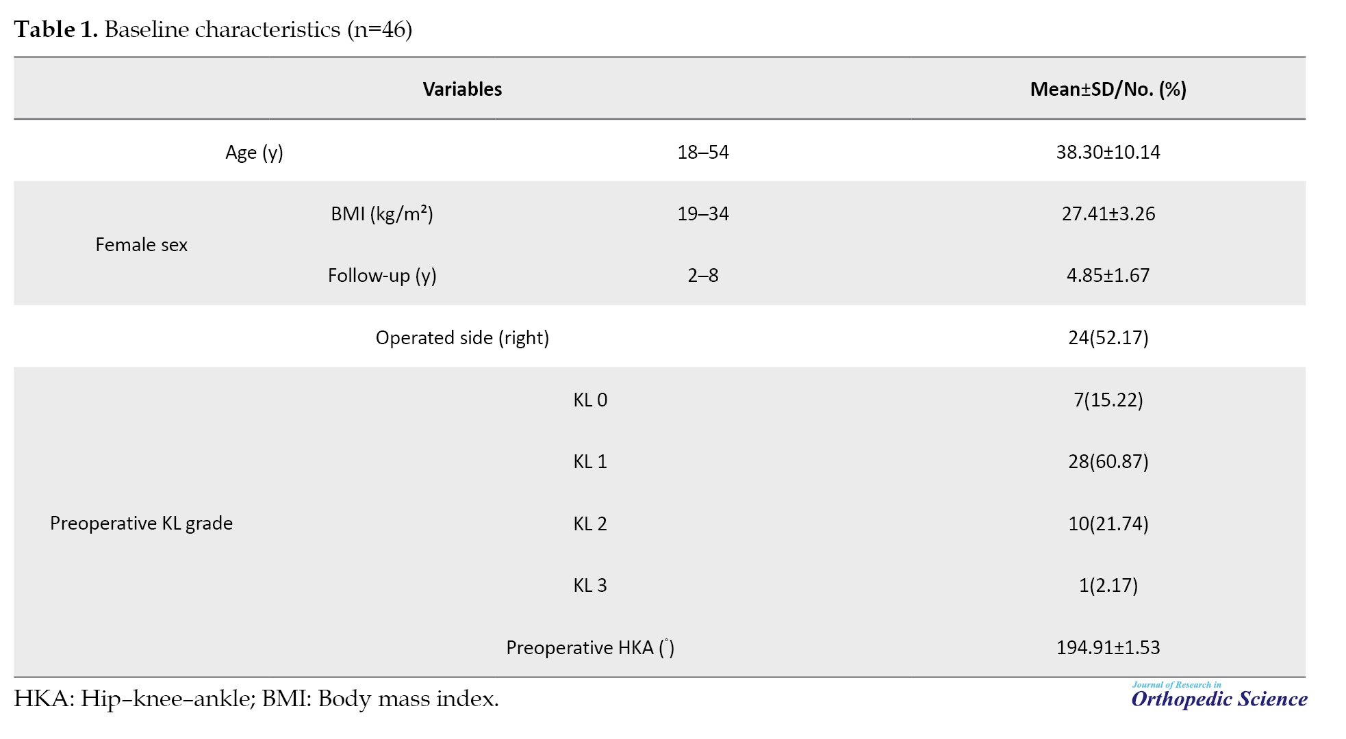
Forty-six patients (46 knees) were included. The mean age was 38.3±10.14 years (range 18–54), and the mean follow-up was 4.85±1.67 years (range 2–8). Thirty-five patients were female (76.09%), and 24 procedures were performed on the right knee (52.17%). Preoperative KL grade was 0 in 7 knees (15.22%), 1 in 28 (60.87%), 2 in 10 (21.74%), and 3 in 1 (2.17%) (Table 1).
Radiographic outcomes
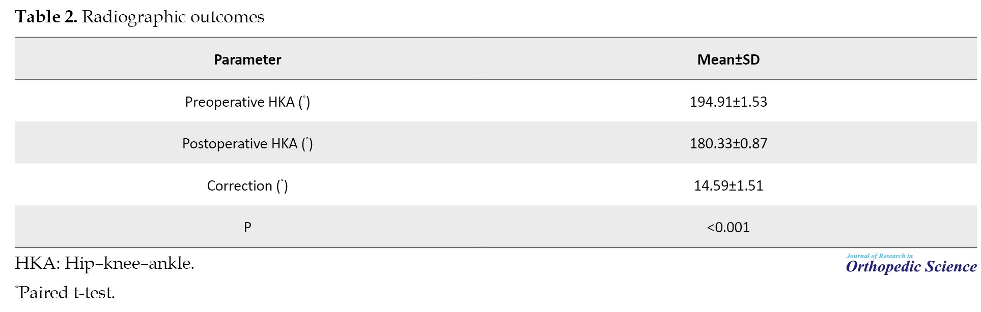
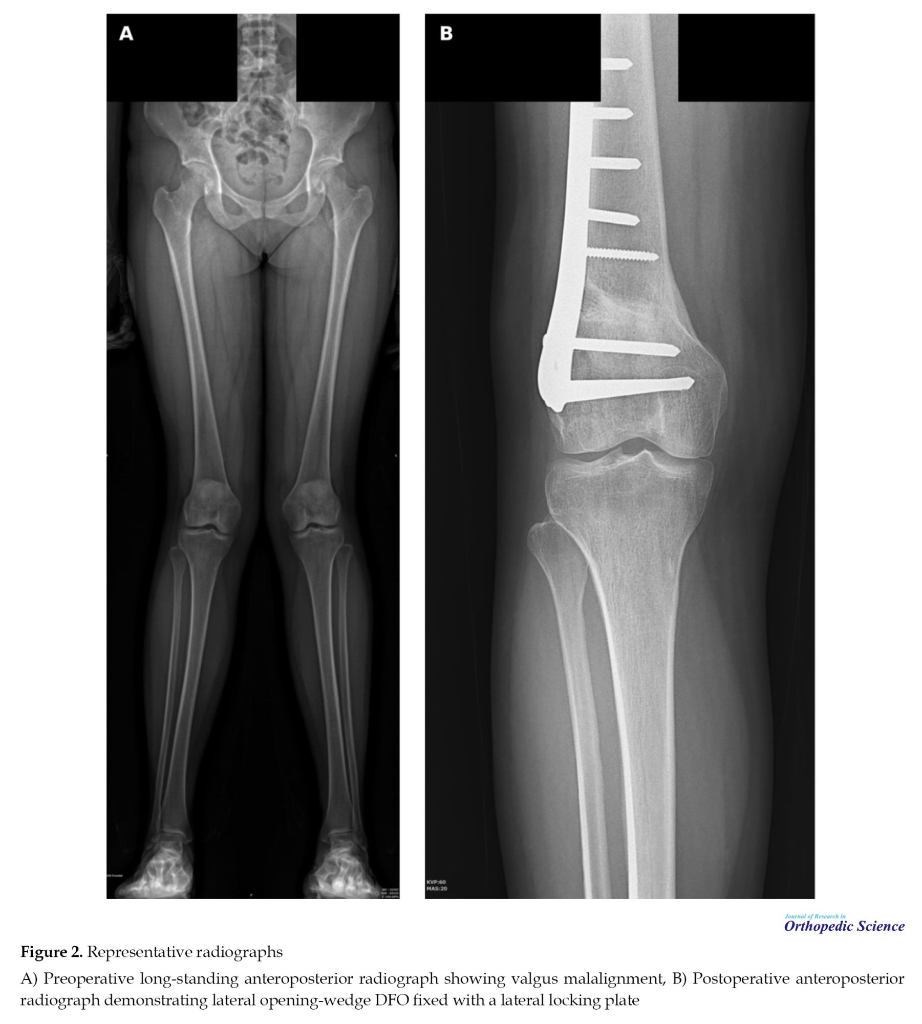
Mean preoperative HKA was 194.91±1.53°, consistent with valgus malalignment. At final follow-up, mean HKA was corrected to 180.33±0.87°, representing a mean correction of 14.59±1.51° (P<0.001) (Table 2, Figure 2).
Clinical outcomes
At final follow-up, the mean VAS pain score was 2.11±1.1, and the mean SF-36 score was 81.59±9.07 (Table 3).
Preoperative patient-reported outcome measures (PROMs) were unavailable, and changes from baseline could not be calculated.
Predictors of outcome
Age correlated negatively with SF-36 (r=-0.36, P=0.01). Higher KL grade was associated with lower SF-36 (Spearman’s rho=-0.50, P<0.001). VAS differed significantly across KL grades (ANOVA, P=0.007) (Figure 3).
Complications and failure
Complications occurred in seven patients (15.22%). Non:union: occurred in six patients (13.04%). One patient (2.17%) underwent conversion to TKA due to persistent pain; this patient had preoperative KL grade 3. Patients with complications were significantly older (P=0.028) and more likely to have KL grade 2 or greater (P=0.046) (Table 4).
Discussion
The principal findings of this retrospective cohort study were that lateral opening-wedge DFO fixed with a locking plate restored near-neutral mechanical alignment with consistent correction and yielded favorable final pain and SF-36 scores at a mean follow-up of 4.85 years. Conversion to TKA occurred in one patient (2.17%), while non:union: was the predominant complication (13.04%). Older age and more advanced radiographic osteoarthritis (KL grade) were associated with worse outcomes and an increased risk of complications.
Radiographic correction is central to the rationale of DFO because shifting the mechanical axis toward neutral unloads the lateral compartment and is the core premise of joint-preserving treatment. In our cohort, mean HKA improved from 194.91° to 180.33°, with a postoperative SD of 0.87°, suggesting a high degree of correction consistency. Systematic reviews and meta-analyses have similarly shown that both opening- and closing-wedge DFO can achieve correction to neutral or slight varus when deformity correction is performed at the origin, and stable fixation is used [3–5]. Studies evaluating correction accuracy after lateral opening-wedge DFO with locking plate constructs have also reported high accuracy, supporting the reliability of contemporary fixation [14].
The final PROMs in this series were favorable (VAS 2.11; SF-36 81.59). Baseline PROMs were not available, so improvement from preoperative status could not be quantified. Nevertheless, these final values compare well with other opening-wedge DFO cohorts that report meaningful symptom relief and functional improvement at mid-term follow-up (often using different instruments, such as KOOS) [15–17]. Return to activity is an important patient-centered endpoint, and prior cohorts have reported that most patients resume physical activity and/or return to work and sports after DFO, although participation in high-impact activity may decrease compared with pre-symptomatic levels [18, 19]. Long-term survivorship remains a key outcome for joint-preserving procedures; published series indicate that DFO can delay conversion to arthroplasty in selected patients at 5–10 years, but results vary with age, baseline arthritis severity, and follow-up duration [7, 8, 20, 21].
Complication rates after DFO vary across the literature and depend on technique, fixation, and definitions used. In the present cohort, non:union: was the dominant complication. Non:union: and delayed :union: have been emphasized as specific concerns after lateral opening-wedge DFO, reinforcing the need for careful technique, risk-factor management, and patient counseling regarding potential reoperation and radiographic :union: monitoring [6]. Published technical and clinical series have described strategies to support healing, including stable fixation and, in selected cases, the use of grafts or substitutes to manage the osteotomy gap [22].
We observed clinically intuitive predictors: higher KL grade was associated with worse SF-36 and greater pain, and older age was associated with lower SF-36. These results support emphasizing careful patient selection for joint-preserving surgery, particularly in knees with more advanced radiographic osteoarthritis, and align with prior DFO series and syntheses that baseline disease stage and patient factors influence outcomes [1, 5, 7, 8].
This study has limitations. Its retrospective design may introduce selection and information bias. Baseline PROMs were not available, preventing assessment of improvement and limiting direct comparison with prospective series. The cohort size was modest, and included few patients with advanced radiographic osteoarthritis, limiting conclusions regarding KL grade 3 disease. Finally, we did not analyze additional knee-specific functional scores or activity-level outcomes. Strengths include a homogeneous technique (lateral opening-wedge DFO with locking plate fixation), mid-term follow-up, and complete radiographic alignment data for all included patients.
Conclusion
Lateral opening-wedge DFO fixed with a LCP achieved accurate correction to near-neutral mechanical alignment and favorable final patient-reported outcomes at a mean follow-up of 4.85 years. Conversion to TKA is uncommon, while non:union: remains the most frequent complication. Older age and higher KL grade were associated with worse outcomes and an increased risk of complications.
Ethical Considerations
Compliance with ethical guidelines
The study was approved by the Ethics Committee of Iran University of Medical Sciences, Tehran, Iran (Code: IR.IUMS.REC.1403.1056).
Funding
This research did not receive any grant from funding agencies in the public, commercial, or non-profit sectors.
Authors' contributions
All authors contributed equally to the conception and design of the study, data collection and analysis, interpretation of the results, and drafting of the manuscript. Each author approved the final version of the manuscript for submission.
Conflict of interest
The authors declared no conflict of interest.
References
Valgus malalignment shifts the mechanical axis laterally and increases the contact pressure in the lateral compartment, contributing to pain and progressive degeneration of the lateral compartment. In young and active patients, arthroplasty may be less desirable due to activity restrictions and the risk of future revision. Distal femoral osteotomy (DFO) is a joint-preserving realignment procedure that aims to unload the lateral compartment by restoring a more neutral mechanical axis in knees with primarily femoral deformity [1–5].
DFO can be performed using a medial closing wedge or a lateral opening wedge technique. Systematic reviews suggest both approaches can achieve reliable correction and comparable outcomes when performed with careful preoperative planning and stable fixation; however, complication profiles differ, and delayed :union:/non:union: has been highlighted as a particular concern after lateral opening-wedge techniques [3–6]. Mid- to long-term series report that DFO can delay conversion to arthroplasty in appropriately selected patients, but survivorship and complication rates vary with age, baseline osteoarthritis severity, and treatment of the osteotomy gap [7, 8].
The purposes of this study were (1) to report radiographic correction, final pain and health-related quality of life, and complications after lateral opening-wedge DFO fixed with a locking compression plate (LCP); and (2) to assess factors associated with outcomes and complications. We hypothesized that DFO would restore near-neutral alignment with acceptable final visual analogue scale (VAS) and SF-36 scores, and that older age and higher K grade would be associated with worse outcomes and an increased risk of complications.
Methods
Study design and setting
This retrospective cohort study was conducted at three tertiary referral hospitals. Data were extracted from institutional medical records between October 2016 and October 2024. The study was reported in accordance with the strengthening the reporting of observational studies in epidemiology (STROBE) statement for observational studies (Figure 1) [9].
Participants
Consecutive patients who underwent lateral opening-wedge DFO for symptomatic valgus malalignment were screened. The primary indication was symptomatic valgus alignment on standing long-leg radiographs (hip–knee–ankle [HKA]>180° as measured in our institution) with clinical and/or radiographic evidence of lateral compartment overload. Patients were included if a complete radiographic dataset (pre- and postoperative long-leg radiographs) and final follow-up clinical assessment were available. Patients with inflammatory arthropathy or advanced tricompartmental degeneration were not considered candidates for osteotomy [1, 2].
Surgical technique
All procedures were performed using a lateral opening-wedge DFO via a direct lateral approach, and the DFO was fixed with a lateral LCP. The goal was to restore near-neutral mechanical alignment based on preoperative planning and intraoperative fluoroscopic confirmation.
Postoperative rehabilitation
Range-of-motion exercises were started immediately after surgery as tolerated. Patients remained non-weight-bearing for approximately 6 weeks (1.5 months), followed by progressive loading. Full weight bearing was permitted after approximately 12 weeks (3 months), based on clinical assessment and radiographic review.
Radiographic assessment
Long-leg anteroposterior radiographs were used to evaluate lower-limb alignment. The HKA angle was measured preoperatively and at final follow-up. In this study, values greater than 180 degrees indicated valgus alignment [10]. Preoperative osteoarthritis severity was classified using the Kellgren–Lawrence (KL) grading system on weight-bearing radiographs [11].
Clinical outcomes
Pain at final follow-up was assessed using a VAS, 0–10 [12]. Health-related quality of life at final follow-up was assessed using the 36-item short form health survey (SF-36, 0–100) [13]. Preoperative VAS and SF-36 scores were unavailable in the medical records and were not analyzed.
Complications and failure
Postoperative complications were identified by chart review. In this cohort, complications comprised non:union: and conversion to total knee arthroplasty (TKA). Failure was defined a priori as conversion to arthroplasty (TKA) during follow-up [6].
Statistical analysis
Continuous variables are reported as Mean±SD and range, and categorical variables as counts and percentages. Pre- versus postoperative HKA was compared using a paired t-test. Correlations were assessed using Pearson correlation (age vs SF-36) and Spearman correlation (KL grade vs SF-36). Differences in VAS across KL grades were tested using one-way analysis of variance (ANOVA). Comparisons between patients with and without complications were performed using independent-samples t-tests for continuous variables and Fisher exact tests for categorical variables. Statistical significance was set at P<0.05. Analyses were performed using IBM SPSS software, version 16 (IBM Corp., Armonk, NY, USA).
Use of generative AI tools
Generative AI tools were used only for language refinement during manuscript preparation. All scientific content, analysis, and interpretations were developed and verified by the authors, who take full responsibility for the manuscript.
Results
Cohort characteristics
Forty-six patients (46 knees) were included. The mean age was 38.3±10.14 years (range 18–54), and the mean follow-up was 4.85±1.67 years (range 2–8). Thirty-five patients were female (76.09%), and 24 procedures were performed on the right knee (52.17%). Preoperative KL grade was 0 in 7 knees (15.22%), 1 in 28 (60.87%), 2 in 10 (21.74%), and 3 in 1 (2.17%) (Table 1).
Radiographic outcomes
Mean preoperative HKA was 194.91±1.53°, consistent with valgus malalignment. At final follow-up, mean HKA was corrected to 180.33±0.87°, representing a mean correction of 14.59±1.51° (P<0.001) (Table 2, Figure 2).
Clinical outcomes
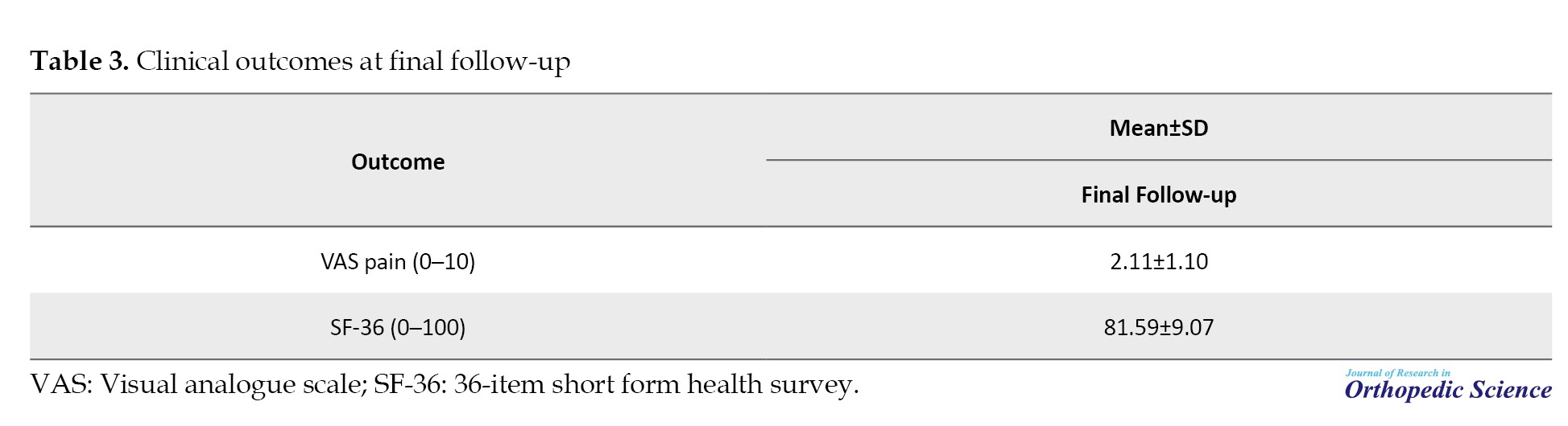
At final follow-up, the mean VAS pain score was 2.11±1.1, and the mean SF-36 score was 81.59±9.07 (Table 3).
Preoperative patient-reported outcome measures (PROMs) were unavailable, and changes from baseline could not be calculated.
Predictors of outcome
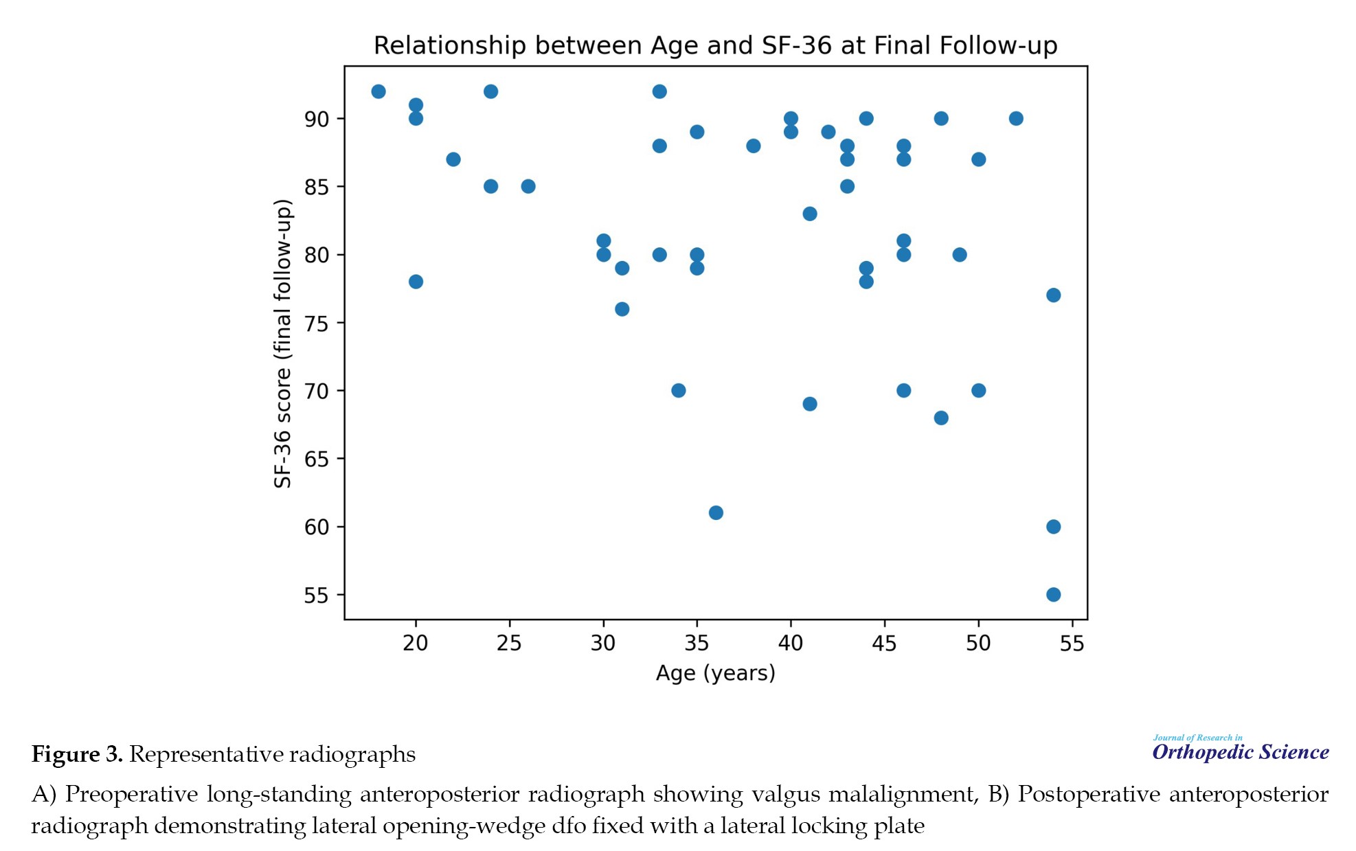
Age correlated negatively with SF-36 (r=-0.36, P=0.01). Higher KL grade was associated with lower SF-36 (Spearman’s rho=-0.50, P<0.001). VAS differed significantly across KL grades (ANOVA, P=0.007) (Figure 3).
Complications and failure
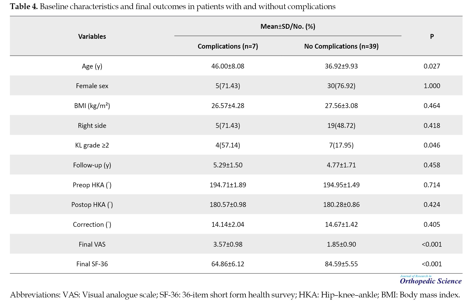
Complications occurred in seven patients (15.22%). Non:union: occurred in six patients (13.04%). One patient (2.17%) underwent conversion to TKA due to persistent pain; this patient had preoperative KL grade 3. Patients with complications were significantly older (P=0.028) and more likely to have KL grade 2 or greater (P=0.046) (Table 4).
Discussion
The principal findings of this retrospective cohort study were that lateral opening-wedge DFO fixed with a locking plate restored near-neutral mechanical alignment with consistent correction and yielded favorable final pain and SF-36 scores at a mean follow-up of 4.85 years. Conversion to TKA occurred in one patient (2.17%), while non:union: was the predominant complication (13.04%). Older age and more advanced radiographic osteoarthritis (KL grade) were associated with worse outcomes and an increased risk of complications.
Radiographic correction is central to the rationale of DFO because shifting the mechanical axis toward neutral unloads the lateral compartment and is the core premise of joint-preserving treatment. In our cohort, mean HKA improved from 194.91° to 180.33°, with a postoperative SD of 0.87°, suggesting a high degree of correction consistency. Systematic reviews and meta-analyses have similarly shown that both opening- and closing-wedge DFO can achieve correction to neutral or slight varus when deformity correction is performed at the origin, and stable fixation is used [3–5]. Studies evaluating correction accuracy after lateral opening-wedge DFO with locking plate constructs have also reported high accuracy, supporting the reliability of contemporary fixation [14].
The final PROMs in this series were favorable (VAS 2.11; SF-36 81.59). Baseline PROMs were not available, so improvement from preoperative status could not be quantified. Nevertheless, these final values compare well with other opening-wedge DFO cohorts that report meaningful symptom relief and functional improvement at mid-term follow-up (often using different instruments, such as KOOS) [15–17]. Return to activity is an important patient-centered endpoint, and prior cohorts have reported that most patients resume physical activity and/or return to work and sports after DFO, although participation in high-impact activity may decrease compared with pre-symptomatic levels [18, 19]. Long-term survivorship remains a key outcome for joint-preserving procedures; published series indicate that DFO can delay conversion to arthroplasty in selected patients at 5–10 years, but results vary with age, baseline arthritis severity, and follow-up duration [7, 8, 20, 21].
Complication rates after DFO vary across the literature and depend on technique, fixation, and definitions used. In the present cohort, non:union: was the dominant complication. Non:union: and delayed :union: have been emphasized as specific concerns after lateral opening-wedge DFO, reinforcing the need for careful technique, risk-factor management, and patient counseling regarding potential reoperation and radiographic :union: monitoring [6]. Published technical and clinical series have described strategies to support healing, including stable fixation and, in selected cases, the use of grafts or substitutes to manage the osteotomy gap [22].
We observed clinically intuitive predictors: higher KL grade was associated with worse SF-36 and greater pain, and older age was associated with lower SF-36. These results support emphasizing careful patient selection for joint-preserving surgery, particularly in knees with more advanced radiographic osteoarthritis, and align with prior DFO series and syntheses that baseline disease stage and patient factors influence outcomes [1, 5, 7, 8].
This study has limitations. Its retrospective design may introduce selection and information bias. Baseline PROMs were not available, preventing assessment of improvement and limiting direct comparison with prospective series. The cohort size was modest, and included few patients with advanced radiographic osteoarthritis, limiting conclusions regarding KL grade 3 disease. Finally, we did not analyze additional knee-specific functional scores or activity-level outcomes. Strengths include a homogeneous technique (lateral opening-wedge DFO with locking plate fixation), mid-term follow-up, and complete radiographic alignment data for all included patients.
Conclusion
Lateral opening-wedge DFO fixed with a LCP achieved accurate correction to near-neutral mechanical alignment and favorable final patient-reported outcomes at a mean follow-up of 4.85 years. Conversion to TKA is uncommon, while non:union: remains the most frequent complication. Older age and higher KL grade were associated with worse outcomes and an increased risk of complications.
Ethical Considerations
Compliance with ethical guidelines
The study was approved by the Ethics Committee of Iran University of Medical Sciences, Tehran, Iran (Code: IR.IUMS.REC.1403.1056).
Funding
This research did not receive any grant from funding agencies in the public, commercial, or non-profit sectors.
Authors' contributions
All authors contributed equally to the conception and design of the study, data collection and analysis, interpretation of the results, and drafting of the manuscript. Each author approved the final version of the manuscript for submission.
Conflict of interest
The authors declared no conflict of interest.
References
- Rosso F, Margheritini F. Distal femoral osteotomy. Curr Rev Musculoskelet Med. 2014; 7(4):302-11. [DOI:10.1007/s12178-014-9233-z] [PMID] [PMCID]
- Puddu G, Cipolla M, Cerullo G, Franco V, Gianni E. Which osteotomy for a valgus knee? Int Orthop. 2010; 34(2):239-47. [DOI:10.1007/s00264-009-0820-3] [PMID] [PMCID]
- Wylie JD, Jones DL, Hartley MK, Kapron AL, Krych AJ, Aoki SK, et al. Distal femoral osteotomy for the valgus knee: medial closing wedge versus lateral opening wedge: A systematic review. Arthroscopy. 2016; 32(10):2141-7. [DOI:10.1016/j.arthro.2016.04.010] [PMID]
- Kim YC, Yang JH, Kim HJ, Tawonsawatruk T, Chang YS, Lee JS, et al. Distal femoral varus osteotomy for valgus arthritis of the knees: Systematic review of open versus closed wedge osteotomy. Knee Surg Relat Res. 2018; 30(1):3-16. [DOI:10.5792/ksrr.16.064] [PMID] [PMCID]
- Diaz CC, Lavoie-Gagne OZ, Knapik DM, Korrapati A, Chahla J, Forsythe B. Outcomes of distal femoral osteotomy for valgus malalignment: A systematic review and meta-analysis of closing wedge versus opening wedge techniques. Am J Sports Med. 2023; 51(3):798-811. [DOI:10.1177/03635465211051740] [PMID]
- Liska F, Voss A, Imhoff FB, Willinger L, Imhoff AB. Non:union: and delayed: :union:: In lateral open wedge distal femoral osteotomies-a legitimate concern? Int Orthop. 2018; 42(1):9-15. [DOI:10.1007/s00264-017-3504-4] [PMID]
- Shivji FS, Foster A, Risebury MJ, Wilson AJ, Yasen SK. Ten-year survival rate of 89% after distal femoral osteotomy surgery for lateral compartment osteoarthritis of the knee. Knee Surg Sports Traumatol Arthrosc. 2021; 29(2):594-9. [DOI:10.1007/s00167-020-05988-5] [PMID]
- Cameron JI, McCauley JC, Kermanshahi AY, Bugbee WD. Lateral opening-wedge distal femoral osteotomy: Pain relief, functional improvement, and survivorship at 5 years. Clin Orthop Relat Res. 2015; 473(6):2009-15. [DOI:10.1007/s11999-014-4106-8] [PMID] [PMCID]
- von Elm E, Altman DG, Egger M, Pocock SJ, Gotzsche PC, Vandenbroucke JP, et al. The strengthening the reporting of observational studies in epidemiology (STROBE) statement: guidelines for reporting observational studies. Plos Med. 2007; 4(10):e296. [DOI:10.1371/journal.pmed.0040296] [PMID] [PMCID]
- Moreland JR, Bassett LW, Hanker GJ. Radiographic analysis of the axial alignment of the lower extremity. J Bone Joint Surg Am. 1987; 69(5):745-9. [DOI:10.2106/00004623-198769050-00016]
- Kellgren JH, Lawrence JS. Radiological assessment of osteoarthrosis. Ann Rheum Dis. 1957; 16(4):494-502. [DOI:10.1136/ard.16.4.494] [PMID] [PMCID]
- Huskisson EC. Measurement of pain. Lancet. 1974; 2(7889):1127-31. [DOI:10.1016/S0140-6736(74)90884-8] [PMID]
- Ware JE Jr, Sherbourne CD. The MOS 36-item short-form health survey (SF-36): I. Conceptual framework and item selection. Med Care. 1992; 30(6):473-83. [DOI:10.1097/00005650-199206000-00002] [PMID]
- Elattar O, Swarup I, Lam A, Nguyen J, Fragomen A, Rozbruch SR. Open wedge distal femoral osteotomy: Accuracy of correction and patient outcomes. HSS J. 2017; 13(2):128-135. [DOI:10.1007/s11420-016-9516-6] [PMID] [PMCID]
- Ekeland A, Nerhus TK, Dimmen S, Heir S. Good functional results of distal femoral opening-wedge osteotomy of knees with lateral osteoarthritis. Knee Surg Sports Traumatol Arthrosc. 2016; 24(5):1702-9. [DOI:10.1007/s00167-016-3988-2] [PMID]
- Kolb A, Isak V, Hobusch GM, Chiari C, Windhager R. Distal femoral varus osteotomy: results of the lateral open-wedge technique without bone grafting. Int Orthop. 2019; 43(10):2315-22. [DOI:10.1007/s00264-018-4216-0] [PMID] [PMCID]
- Saithna A, Kundra R, Getgood A, Spalding T. Opening wedge distal femoral varus osteotomy for lateral compartment osteoarthritis in the valgus knee. Knee. 2014; 21(1):172-5. [DOI:10.1016/j.knee.2013.08.014] [PMID]
- Hoorntje A, van Ginneken BTJ, Kuijer PPFM, Koenraadt KLM, van Geenen RCI, Kerkhoffs GMMJ, et al. Eight respectively nine out of ten patients return to sport and work after distal femoral osteotomy. Knee Surg Sports Traumatol Arthrosc. 2019; 27(7):2345-53. [DOI:10.1007/s00167-018-5206-x] [PMID] [PMCID]
- de Carvalho LH Jr, Temponi EF, Soares LFM, Goncalves MBJ, Costa LP. Physical activity after distal femur osteotomy for the treatment of lateral compartment knee osteoarthritis. Knee Surg Sports Traumatol Arthrosc. 2014; 22(7):1607-11. [DOI:10.1007/s00167-012-2316-8] [PMID]
- McDermott AG, Finkelstein JA, Farine I, Boynton EL, MacIntosh DL, Gross A. Distal femoral varus osteotomy for valgus deformity of the knee. J Bone Joint Surg Am. 1988; 70(1):110-6. [DOI:10.2106/00004623-198870010-00017]
- Aglietti P, Buzzi R, Vena LM, Baldini A, Mondaini A. Distal femoral varus osteotomy in valgus osteoarthritis of the knee. Am J Knee Surg. 2000; 13(2):89-95. [Link]
- Dewilde TR, Dauw J, Vandenneucker H, Bellemans J. Opening wedge distal femoral varus osteotomy using the Puddu plate and calcium phosphate bone cement. Knee Surg Sports Traumatol Arthrosc. 2013; 21(1):249-54. [DOI:10.1007/s00167-012-2156-6] [PMID]
Type of Study: Research Article |
Subject:
Knee surgery
Received: 2025/08/22 | Accepted: 2026/02/1 | Published: 2026/04/1
Received: 2025/08/22 | Accepted: 2026/02/1 | Published: 2026/04/1
| Rights and permissions | |
 |
This work is licensed under a Creative Commons Attribution-NonCommercial 4.0 International License. |

Copyright © The Author(s);
This is an open access article distributed under the terms of the Creative Commons Attribution License (CC-By-NC), which permits use, distribution, and reproduction in any medium, provided the original work is properly cited and is not used for commercial purposes.
Contact Information
