

Volume 13, Issue 1 (Winter 2026)
JROS 2026, 13(1): 13-24 |
Back to browse issues page




Download citation:
BibTeX | RIS | EndNote | Medlars | ProCite | Reference Manager | RefWorks
Send citation to:


BibTeX | RIS | EndNote | Medlars | ProCite | Reference Manager | RefWorks
Send citation to:
Moghtadaei M, Farahini H, Yegane A, Ameri M, Zarei B, Sour B. Intramedullary Versus Extramedullary Tibial Alignment Guides in Total Knee Arthroplasty: Coronal Alignment and Early Inflammatory Marker Trends. JROS 2026; 13 (1) :13-24
URL: http://jros.iums.ac.ir/article-1-2323-en.html
URL: http://jros.iums.ac.ir/article-1-2323-en.html


1- Department of Orthopedic Surgery, School of Medicine, Iran University of Medical Sciences, Tehran, Iran.
2- Clinical Research Development Center, Rasoul Akram Hospital Complex, Iran University of Medical Sciences, Tehran, Iran.
3- Bone and Joint Reconstruction Research Center, Iran University of Medical Sciences, Tehran, Iran.
2- Clinical Research Development Center, Rasoul Akram Hospital Complex, Iran University of Medical Sciences, Tehran, Iran.
3- Bone and Joint Reconstruction Research Center, Iran University of Medical Sciences, Tehran, Iran.
Keywords: Arthroplasty, Replacement, Knee, Tibial alignment, Intramedullary (IM), Extramedullary (EM), Inflammatory markers
Full-Text [PDF 1089 kb]
(32 Downloads)
| Abstract (HTML) (635 Views)
Full-Text: (3 Views)
Introduction
Restoration of coronal limb alignment has traditionally been considered a key objective in total knee arthroplasty (TKA), given reported associations with implant longevity and biomechanical load distribution [1-3]. Malalignment of the mechanical axis has been linked to increased wear and higher revision rates in long-term follow-up series [1, 3]. Therefore, accurate tibial component positioning is central to achieving an appropriate postoperative mechanical axis.
Intramedullary (IM) and extramedullary (EM) tibial alignment guides are widely used for tibial resection in conventional TKA. IM guides reference the tibial canal, whereas EM guides rely on external anatomical landmarks. Comparative series and randomized trials have often reported similar coronal alignment accuracy and comparable outlier rates between techniques [4-12]. Systematic reviews and meta-analyses generally suggest that no consistent alignment advantage exists for either guide across heterogeneous settings [13-16].
In addition to alignment, the degree of IM instrumentation may influence perioperative physiology through canal violation and endosteal trauma. Indirect evidence from studies comparing conventional instrumentation with approaches that avoid IM rods (for example, robotic or navigated procedures) suggests that differences in systemic inflammatory markers may be modest and transient [17, 18]. Direct data specifically comparing inflammatory marker kinetics between IM and EM tibial alignment techniques are limited.
Objectives
This study aimed to compare coronal alignment parameters and perioperative inflammatory marker trends between IM and EM tibial alignment techniques using real-world clinical data collected during routine care. This dual-domain evaluation was intended to assess both mechanical alignment and early systemic biological responses in the same cohort. We hypothesized that alignment outcomes would be similar between techniques and that any differences in systemic inflammatory response would be small.
Methods
Study design and data sources
This prospective pragmatic observational cohort study used clinical, laboratory, and radiographic data collected during routine perioperative care. All clinical and laboratory variables were recorded prospectively from routine clinical documentation. No additional imaging was obtained for research purposes.
The study protocol was registered in an institutional research management system (tracking code: blinded for review; registered on February 7, 2025).
This study was conducted in accordance with the ethical guidelines of the 1975 Declaration of Helsinki, and written informed consent was obtained from each patient included in the study.
Setting and recruitment
This prospective, pragmatic, observational cohort study was conducted across three tertiary orthopedic centers in 2025. Consecutive patients scheduled for primary TKA were screened for eligibility. Clinical, radiographic, and laboratory data were collected prospectively according to routine care pathways at predefined perioperative time points. Alignment radiographs were obtained as part of standard postoperative assessment.
Eligibility criteria
The inclusion criteria included adults undergoing primary TKA for degenerative knee disease, use of either an IM or EM tibial alignment guide, and availability of standing long-leg radiographs preoperatively and/or postoperatively as part of routine care. The exclusion criteria included revision TKA, prior tibial osteotomy, severe extra-articular deformity requiring additional corrective procedures, and inflammatory arthropathy or active infection at the time of surgery. Patients with incomplete laboratory follow-up were retained; missing laboratory values were handled by available-case analysis.
Participants and grouping
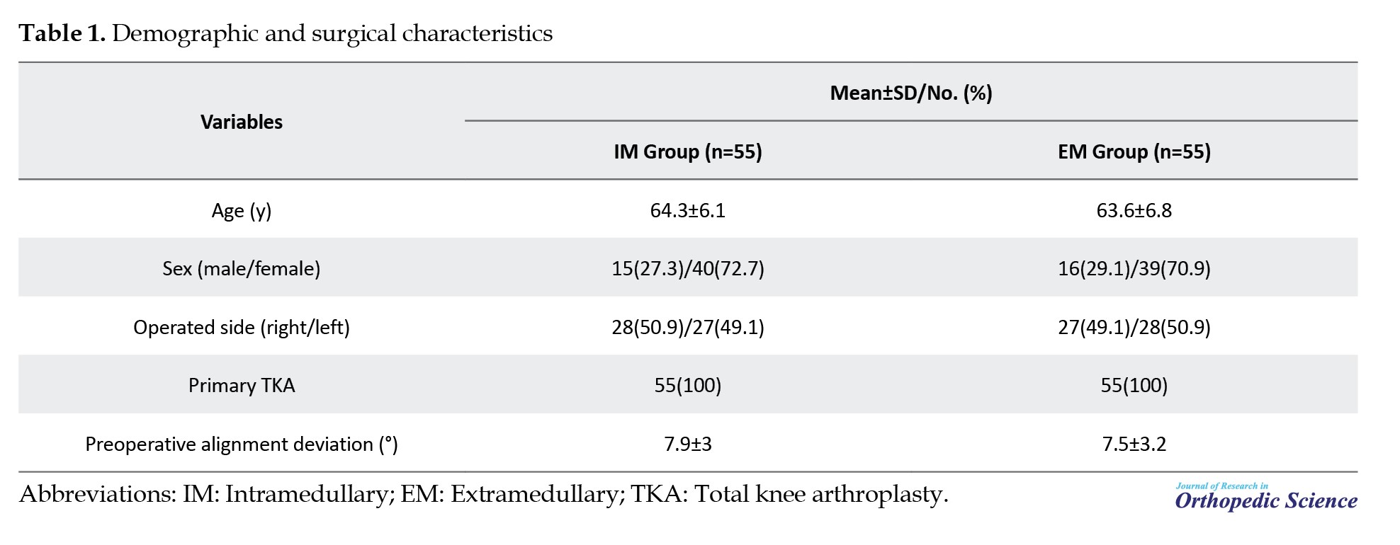
A total of 110 primary TKA cases were enrolled and analyzed. The cases were grouped by the tibial alignment technique used during surgery: IM (IM group, n=55) or EM (EM group, n=55). Use of IM versus EM tibial alignment guides was determined by surgeon preference and intraoperative feasibility; allocation was non-random. Table 1 presents demographic and baseline surgical characteristics.
Exposure definition (tibial alignment technique)
The tibial alignment method used during surgery was the focus of the exposure. In the IM group, tibial alignment was guided by an IM rod inserted into the tibial canal according to the manufacturer’s technique. In the EM group, tibial alignment was guided by an EM jig referenced to external anatomical landmarks. The selection of IM versus EM guidance was non-random and determined by surgeon preference and intraoperative feasibility.
Outcomes
The primary outcomes were coronal alignment parameters on standing long-leg radiographs (hip-knee-ankle [HKA], lateral distal femoral angle [LDFA], medial proximal tibial angle [MPTA], intramedullary alignment angle [IMA], and joint line convergence angle [JLCA], when available and perioperative inflammatory marker trajectories (white blood cell count [WBC], erythrocyte sedimentation rate [ESR], C-reactive protein [CRP], interleukin-6 [IL-6]). The secondary outcome was the proportion of alignment outliers, defined as an absolute HKA deviation >3° from neutral” consistently. Outcomes were assessed using routinely collected clinical data and imaging without protocol-driven additional testing.
Study size
The study size was determined based on the number of eligible cases accrued during the study period. No formal a priori sample size calculation was performed because the study was designed to describe real-world variability in alignment and perioperative laboratory kinetics rather than to detect small between-group differences with high precision.
Perioperative management
All patients were managed using standardized institutional perioperative pathways, including antibiotic prophylaxis, routine thromboprophylaxis, and postoperative rehabilitation protocols. Blood sampling for inflammatory markers was performed as part of routine postoperative monitoring at predefined time points. The surgical approach and implant type were performed according to standard practice at each center. Tourniquet use, tranexamic acid administration, and drain placement were performed according to surgeon preference; these variables were not controlled for in the study protocol.
Radiographic measurements
Standing full-length lower-limb radiographs were reviewed. Alignment angles were extracted from radiology reports when documented as numeric values. When angles were not reported numerically, radiographic measurements were independently performed by two trained reviewers using standardized coronal-plane mechanical-axis techniques. Both reviewers were blinded to the surgical technique and laboratory outcomes. Measurements were conducted in a picture archiving and communication system (PACS) using calibrated digital angle tools according to a predefined protocol. In cases of discrepancy, a final consensus measurement was obtained through joint review. Radiographic parameters included HKA angle, LDFA, MPTA, IMA, and JLCA when available. Alignment outliers were defined a priori as deviations >±3° from neutral mechanical alignment (HKA=180°).
Angles were defined using standard coronal plane conventions: HKA, the coronal angle between the femoral and tibial mechanical axes; LDFA, the lateral angle between the femoral mechanical axis and the distal femoral joint line; MPTA, the medial angle between the tibial mechanical axis and the proximal tibial joint line; and JLCA, the angle between the distal femoral and proximal tibial joint lines. Measurements were recorded in degrees as documented or measured in PACS.
Inflammatory markers
The perioperative inflammatory markers included WBC, ESR, CRP, and IL-6. Values were recorded preoperatively and on postoperative days 1, 7, and 15, with CRP additionally recorded on postoperative day 3 to capture the typical postoperative peak, when available as part of routine clinical care.
Missing data
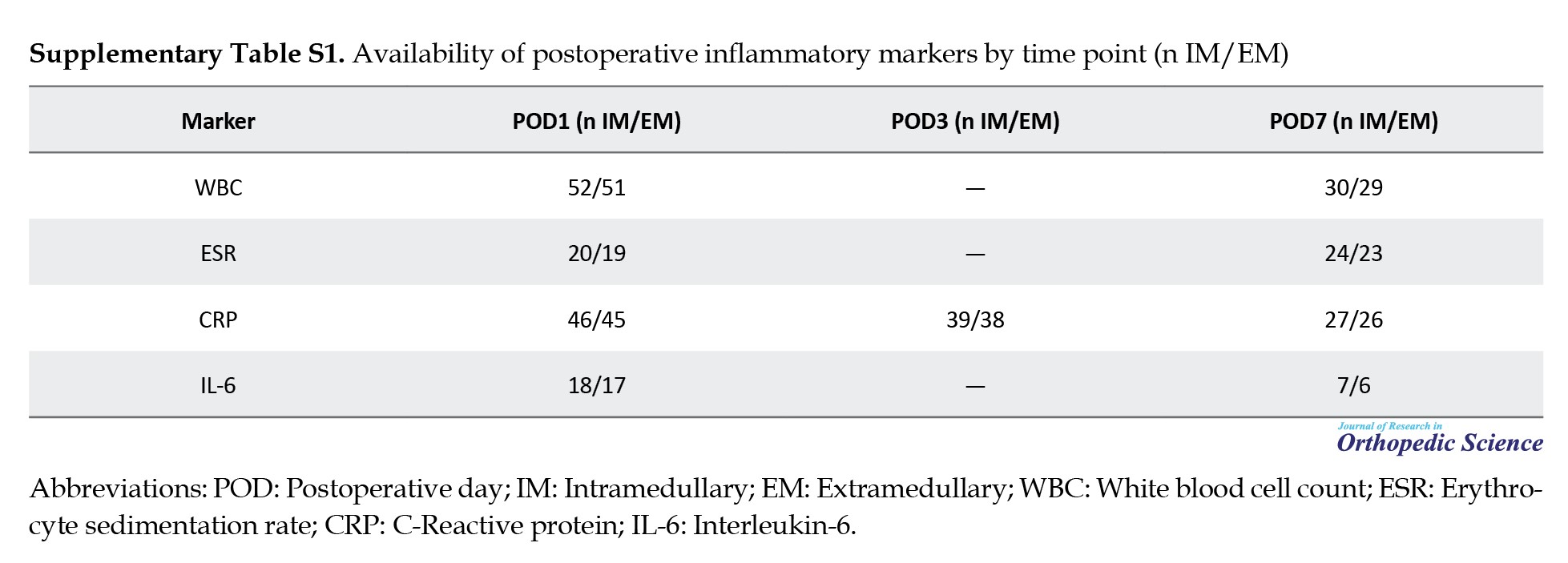
Laboratory markers were obtained as part of routine care; therefore, availability varied by time point. Available case analysis was performed without imputation. The number of observations per group at each postoperative time point is reported for each marker to support interpretability and avoid overstating precision (Supplementary Supplementary Table S1).
Because laboratory sampling was not fully standardized, inflammatory-marker analyses were interpreted as exploratory.
Potential sources of bias
Several steps were taken to reduce bias within the constraints of routine care data. IM versus EM guidance was recorded from operative documentation, and laboratory outcomes were abstracted directly from institutional results. Radiographic angles were obtained from reports when available, and PACS measurements were performed by two trained reviewers using a predefined standard protocol with blinding to surgical technique and laboratory results. Residual confounding remains possible because exposure assignment was non-random and factors such as comorbidities, operative time, tourniquet use, and tranexamic acid use were not controlled for in the protocol.
Statistical analysis
Continuous variables are presented as Mean±SD and categorical variables as No. (%). Between-group comparisons at each time point were assessed using Welch’s t-test for continuous variables and Fisher’s exact test for categorical variables, including alignment outliers. Mean differences with 95% confidence intervals (CI) were calculated using Welch standard errors. For binary outcomes, risk differences with 95% confidence intervals (CIs) were reported. Analyses were two-sided; P<0.05 was considered statistically significant. Given multiple time points and outcomes, P values were interpreted as exploratory and were not adjusted for multiple comparisons. No multivariable adjustment was performed because the study was designed as an exploratory routine-care cohort and covariate completeness was limited across outcomes.
This study was reported in accordance with the strengthening the reporting of observational studies in epidemiology (STROBE) statement.
Results
Participant flow and availability
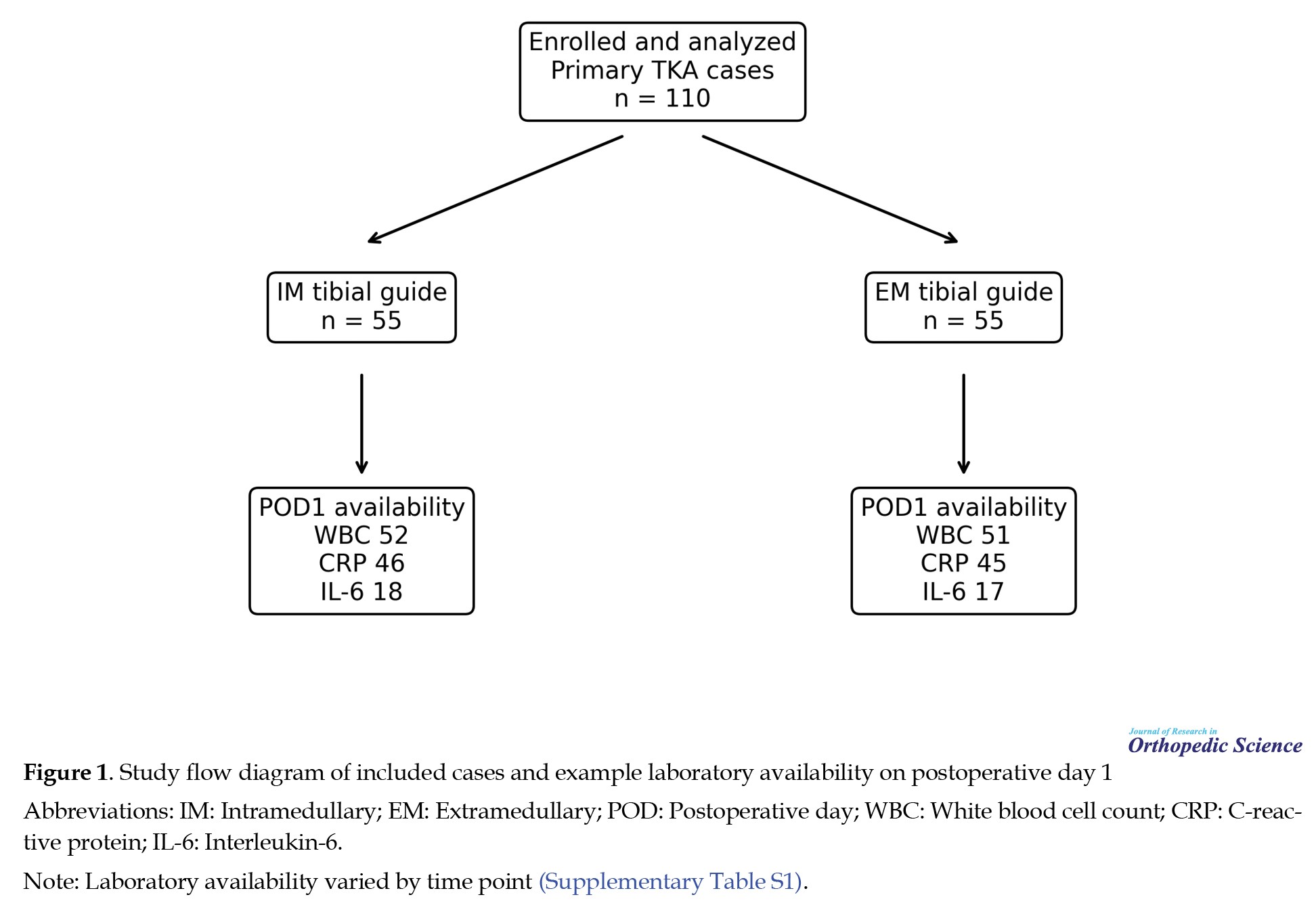
A total of 110 patients were enrolled and analyzed (55 IM, 55 EM); all were primary TKAs. Postoperative laboratory availability decreased over time, consistent with routine follow-up patterns. For example, WBC was available for 52/51 patients on postoperative day 1 and 30/29 patients on postoperative day 7 (IM/EM). IL-6 measurements were available in smaller subsets (18/17 on day 1; 7/6 on day 7). Radiographic parameters were available for a subset depending on documentation and imaging availability. Figure 1 shows the participant flow and the availability of postoperative laboratory results.
Patient characteristics
The study cohort comprised 110 primary TKA cases (55 IM and 55 EM). The mean age was 64.3±6.1 years in the IM group and 63.6±6.8 years in the EM group (P=0.571). The sex distribution was similar (IM: 15 males/40 females; EM: 16 males/39 females; P=1.000), as was the operated side (IM: 28 right/27 left; EM: 27 right/28 left; P=1.000) (Table 1). The mean preoperative alignment deviation was 7.9±3.0° in the IM group and 7.5°±3.2° in the EM group (P=0.500). All cases were primary TKA.
Radiographic coronal alignment
Documented coronal alignment parameters showed similar mean point estimates between groups (Table 2).
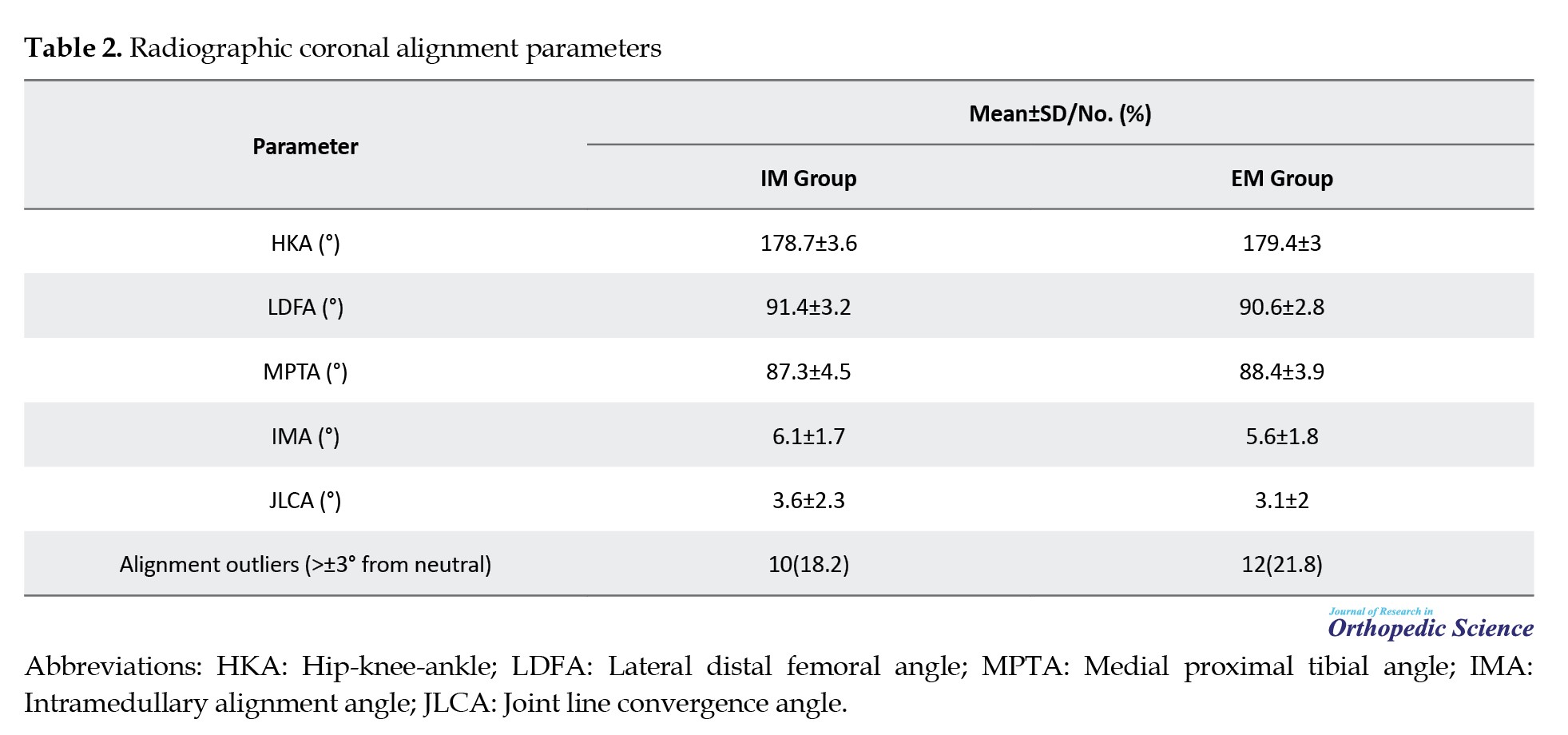
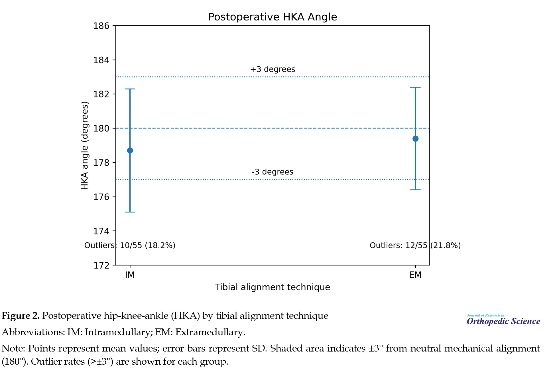
The mean HKA was 178.7±3.6° in the IM group and 179.4±3.0° in the EM group (mean difference -0.7°, P=0.270). No evidence of a clear between-group difference was observed for mean LDFA (P=0.166), MPTA (P=0.174), IMA (P=0.137), or JLCA (P=0.226). Alignment outliers (absolute HKA deviation >3° from neutral) occurred in 10/55 (18.2%) IM cases and 12/55 (21.8%) EM cases (P=0.812). The risk difference was -3.6 percentage points (95% confidence interval [CI] -18.6 to 11.3; P=0.812). Figure 2 shows mean postoperative HKA values and the ±3° threshold.
Perioperative inflammatory markers
All inflammatory markers increased after surgery and demonstrated expected postoperative kinetics (Tables 3, 4, 5, and 6).
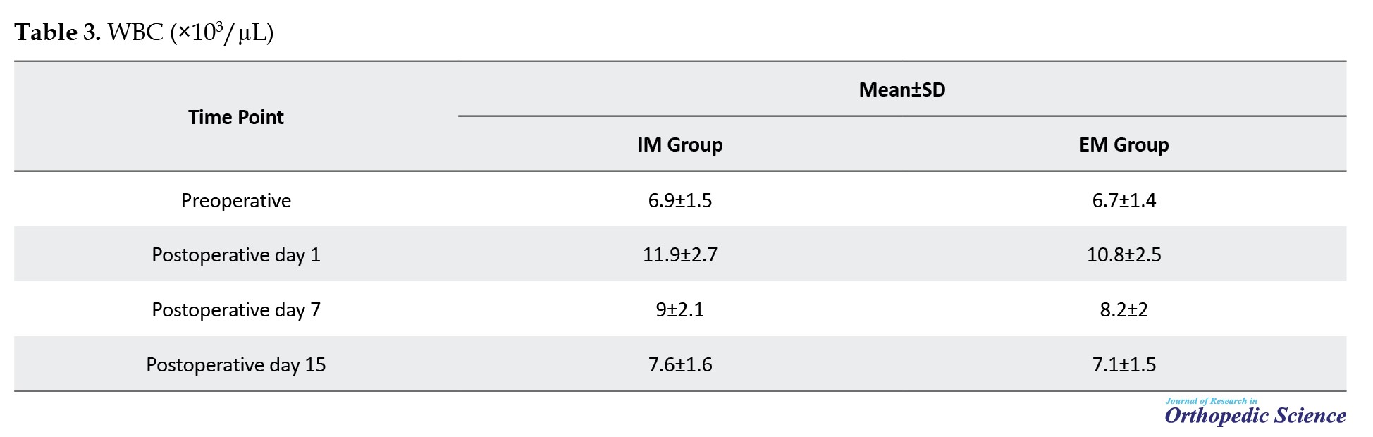
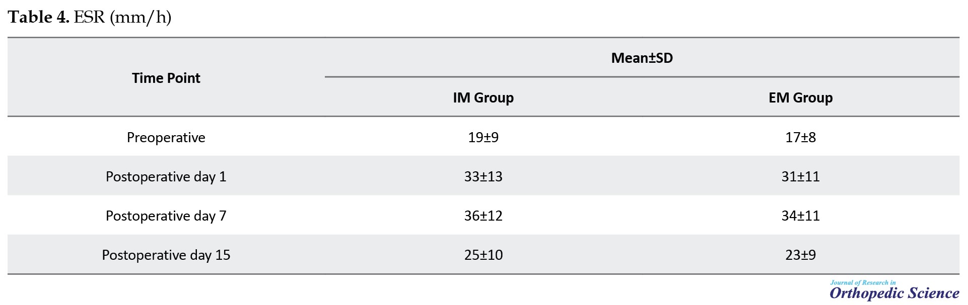
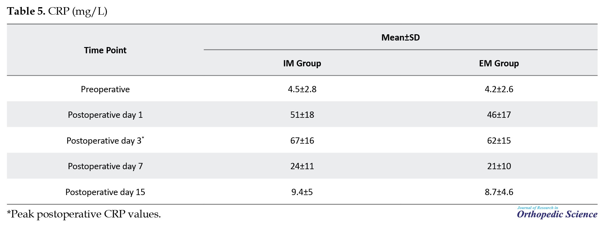
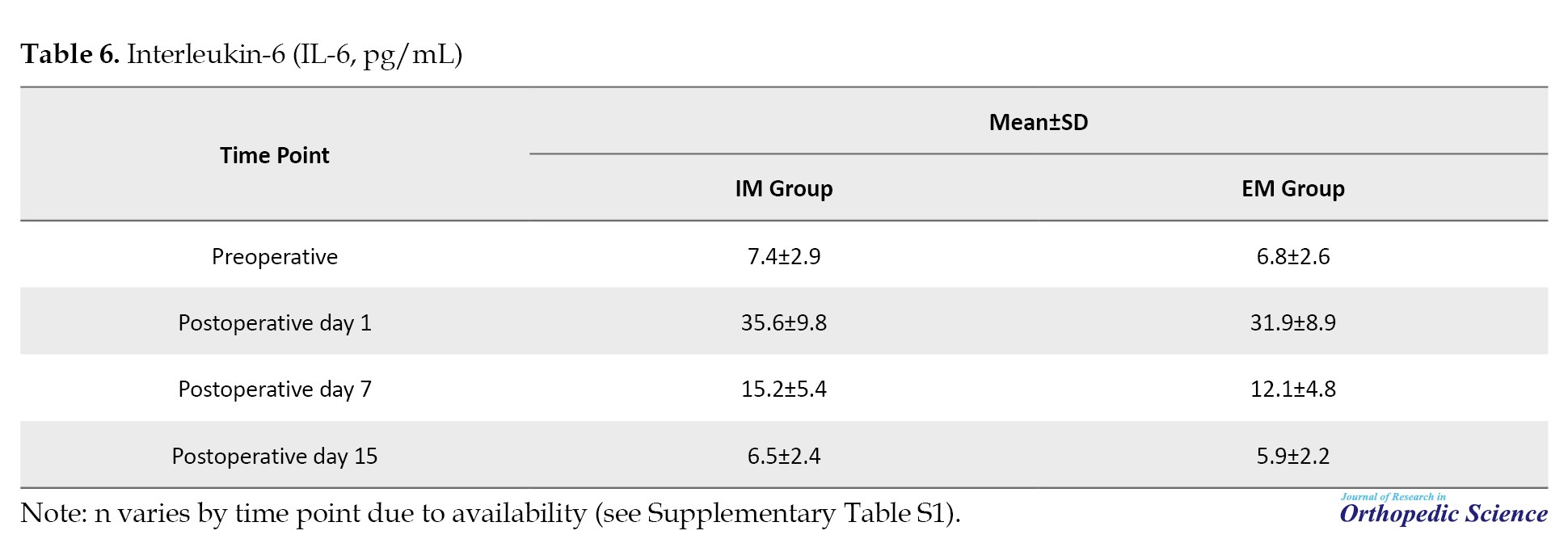
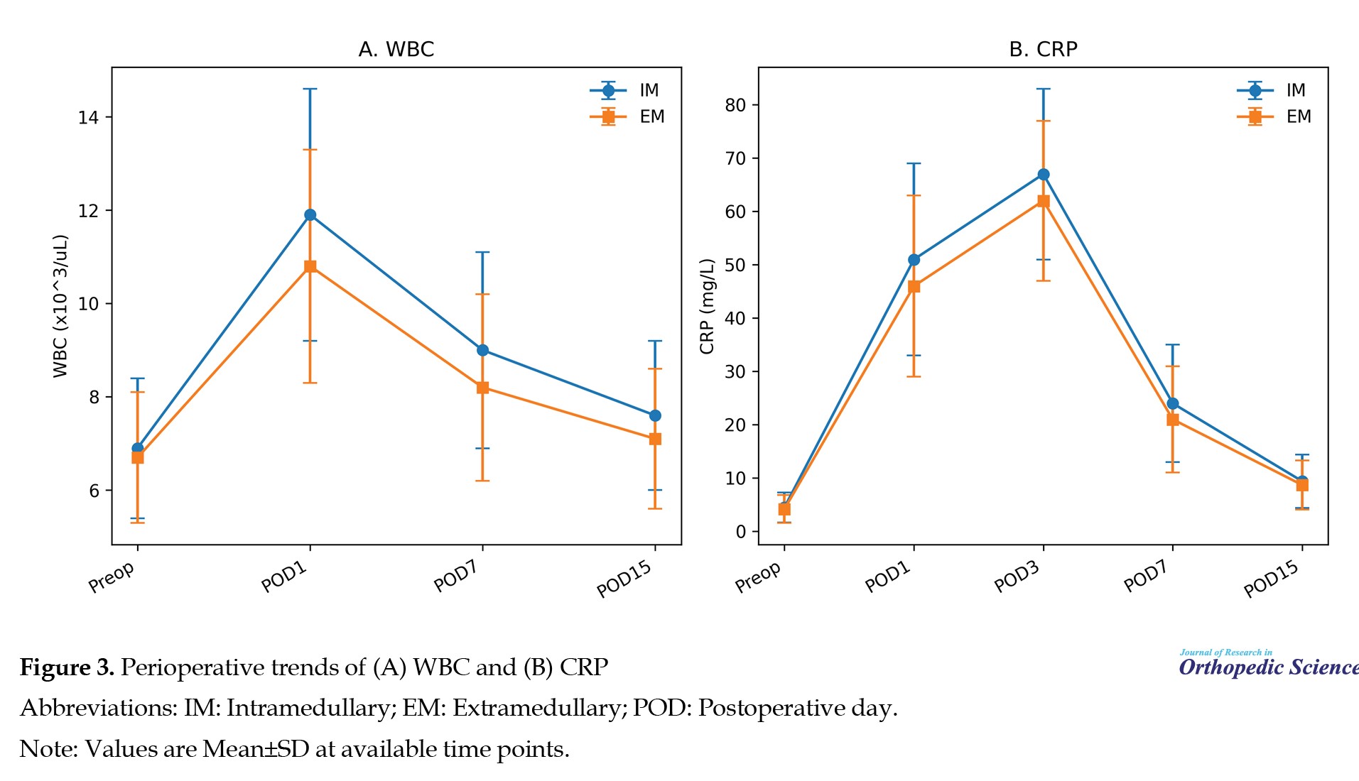
The available sample sizes varied by marker and time point (Supplementary Table S1). WBC was modestly higher in the IM group on postoperative day 1 (11.9±2.7 vs 10.8±2.5×10³/µL; mean difference 1.1, 95% CI 0.08 to 2.12; P=0.034). This isolated finding should be interpreted cautiously because multiple exploratory comparisons were performed. No evidence of a clear between-group difference was observed at later WBC time points (postoperative day 7: P=0.139). CRP peaked on postoperative day 3 in both groups (67±16 vs 62±15 mg/L; mean difference 5, 95% CI -2.0 to 12.0; P=0.161). IL-6 comparisons were limited by the small number of available samples and showed no clear difference at the reported time points (postoperative day 1: P=0.250; postoperative day 7: P=0.297). WBC peaked on postoperative day 1 and declined toward baseline by day 15 in both groups. ESR rose more gradually and remained elevated at day 15. IL-6 peaked early and declined by postoperative day 15. Figure 3 shows WBC and CRP trajectories.
Discussion
The principal finding of this prospective pragmatic observational cohort study was that the point estimates for coronal alignment were similar between the IM and EM tibial guide groups in routine clinical practice. No evidence of a clear between-group difference was observed for HKA, LDFA, MPTA, IMA, or JLCA in the available sample. The main contribution of this study is the combined assessment of radiographic alignment and early perioperative inflammatory marker trends between IM and EM tibial alignment techniques in routine clinical practice.
Comparison with existing literature
Our alignment findings are consistent with most comparative evidence, suggesting broadly similar coronal alignment accuracy for IM and EM tibial guides. Randomized trials and meta-analyses have reported no meaningful difference in postoperative mechanical axis or tibial component coronal alignment between techniques, with comparable pooled outlier rates [4, 8, 10, 13-16]. A randomized trial in severely obese patients also reported equivalent tibial alignment using IM versus EM guidance, supporting applicability in challenging anatomy [8]. However, individual studies have reported context-dependent differences; for example, Razzaghof et al. observed fewer MPTA outliers with IM guidance in their randomized trial [19]. Taken together, these findings suggest that small differences in outlier rates may reflect patient anatomy, surgeon technique, and landmark reliability rather than a consistent intrinsic advantage of one guide.
Compared with single-surgeon randomized trials, our real-world cohort likely captures greater variability in workflow and documentation. This may contribute to a wider spread of alignment values; however, our outlier rate remained within the range reported in controlled studies and pooled analyses. These observations support the pragmatic interpretation that either conventional guide can achieve acceptable coronal alignment when applied carefully, and that surgeon familiarity and intraoperative judgment remain central determinants of alignment.
Several factors may influence the practical performance of EM versus IM guidance. External landmarking may be challenged by soft-tissue envelope, ankle deformity, or rotational positioning, whereas IM referencing may be affected by canal anatomy, bowing, or prior deformity. Accordingly, studies have reported variable patterns of outliers, with some suggesting fewer tibial coronal outliers with IM guidance in selected settings, while others show equivalence. Our findings support a conservative interpretation that guide choice alone is unlikely to be the dominant determinant of coronal alignment.
Beyond alignment, some authors have examined whether IM instrumentation influences perioperative blood loss or physiological stress. A large retrospective series comparing intra- versus EM alignment approaches reported similar estimated blood loss and transfusion rates, suggesting that canal entry alone may not translate into clinically meaningful differences in bleeding in modern practice [20]. A recent single-centre cohort also reported satisfactory radiographic and perioperative outcomes with IM tibial guidance [21]. Indirect comparisons from navigation or robotic-assisted TKA workflows that may reduce IM rod use have reported modest short-term differences in systemic biomarkers. However, these effects were transient and may be influenced by multiple aspects of technique rather than a single instrument choice [17, 18]. In the present study, laboratory trajectories were broadly similar between tibial guide groups, supporting the view that any systemic impact of tibial alignment method is likely small relative to the overall surgical insult; however, the laboratory findings should be considered exploratory because sampling was incomplete and not fully standardized.
Perioperative inflammatory response in context
Early postoperative systemic inflammatory marker trajectories were broadly similar between groups. WBC was modestly higher in the IM group on postoperative day 1. In contrast, no evidence of a clear difference was observed in other available inflammatory marker comparisons, and IL-6 analyses were limited by the small sample size. These findings are biologically plausible given the hypothesis that IM canal violation may transiently influence systemic physiology; however, causal inference is limited by the non-randomized design, lack of multivariable adjustment, and incomplete laboratory capture. Indirect evidence from studies avoiding IM instrumentation (e.g. robotic or navigated workflows) suggests modest, short-lived reductions in early inflammatory markers compared with conventional techniques [17, 18].
The within-group trajectories observed in this cohort were consistent with established postoperative kinetics after uncomplicated TKA, including an early rise in IL-6 and a CRP peak around postoperative days 2-3 with subsequent decline [22, 23], while ESR showed a more prolonged elevation in our cohort. In our data, between-group differences were small relative to the magnitude of the expected postoperative response, and confidence intervals for key comparisons were compatible with both no effect and modest effects. Together, this suggests that any systemic inflammatory impact of tibial guide selection is likely limited in typical primary TKA, although larger studies with standardized sampling are needed for definitive inference.
Clinical implications
In routine primary TKA, guide selection should be individualized. Based on our findings and the broader literature, practical considerations include:
EM guidance may be preferred when tibial canal access is undesirable or IM access is limited by anatomy or prior instrumentation.
IM guidance may be helpful when external landmarks are unreliable (e.g. high soft-tissue envelope), although evidence for reduced coronal outliers is inconsistent across studies.
Surgeon familiarity, careful landmarking, and intraoperative verification likely have a greater impact on alignment than guide type alone.
Clinical significance should be interpreted cautiously. We did not evaluate clinical outcomes, complications, or infection endpoints in relation to inflammatory marker trajectories, and residual confounding, including operative time, tissue handling, tourniquet use, tranexamic acid use, and comorbidity profiles, may influence inflammatory response. Multiple comparisons were performed across time points; P values should be considered exploratory.
The strengths of the study include prospective data capture in routine practice, inclusion of consecutive primary TKA cases, standardized two-reviewer radiographic measurement when report values were unavailable, and combined evaluation of radiographic alignment and perioperative laboratory markers. Reporting of availability and effect estimates was intended to improve transparency in the setting of real-world missingness.
Conclusion
In this prospective pragmatic observational cohort, IM and EM tibial alignment techniques in TKA showed similar coronal alignment as assessed by standing long-leg radiographs. Perioperative inflammatory marker trajectories were broadly similar in available samples, with a modestly higher WBC on postoperative day 1 in the IM group. Because of the non-randomized design and incomplete routine-care laboratory sampling, these findings should be interpreted as exploratory. Further studies with standardized measurement protocols, more complete laboratory data capture, adjusted analyses, and correlation with clinical outcome are warranted.
Limitations
This study has limitations inherent to its observational design. The allocation of IM versus EM guidance was non-random, introducing potential for unmeasured confounding and selection bias due to surgeon preference. No multivariable adjustment was performed, and variables, such as operative time, tourniquet use, tranexamic acid use, tissue handling, and comorbidity profiles were not controlled. Alignment parameters were partly report-derived, and a subset required PACS-based measurement from existing radiographs, which may introduce measurement heterogeneity. Although measurements were performed by two trained reviewers using a standardized protocol, formal interobserver and intraobserver reliability testing was not performed. Laboratory testing schedules were not uniform across patients, reducing power for some comparisons, particularly IL-6, and limiting inference; missing data were not imputed. Clinical outcomes, complications, and infection endpoints were not assessed.
Ethical Considerations
Compliance with ethical guidelines
This study was approved by the Research Ethics Committee of Iran University of Medical Sciences, Tehran, Iran (Code: IR.IUMS.REC.1404.566). The study protocol was registered in the institutional research management system (tracking code: blinded for review). The study was conducted in accordance with the Declaration of Helsinki. Written informed consent was obtained from all participants. The submitted manuscript contains no names, initials, hospital identification numbers, or photographs that could identify patients.
Funding
This research did not receive any grant from funding agencies in the public, commercial, or non-profit sectors.
Authors' contributions
Conceptualization and methodology: Mehdi Moghtadaei, Hossein Farahini, Ali Yegane, and Mahmoud Ameri; Data collection: Mehdi Moghtadaei, Hossein Farahini, and Bushra Zarei; Formal analysis: Ali Yegane and Bushra Zarei; Analysis: Mahmoud Ameri and Ali Yegane; Writing the original draft: Mahmoud Ameri; Review and editing: Behnam Sour; Supervision: Mehdi Moghtadaei and Hossein Farahini; Final approval: All authors.
Conflict of interest
The authors declared no conflict of interest.
References
Restoration of coronal limb alignment has traditionally been considered a key objective in total knee arthroplasty (TKA), given reported associations with implant longevity and biomechanical load distribution [1-3]. Malalignment of the mechanical axis has been linked to increased wear and higher revision rates in long-term follow-up series [1, 3]. Therefore, accurate tibial component positioning is central to achieving an appropriate postoperative mechanical axis.
Intramedullary (IM) and extramedullary (EM) tibial alignment guides are widely used for tibial resection in conventional TKA. IM guides reference the tibial canal, whereas EM guides rely on external anatomical landmarks. Comparative series and randomized trials have often reported similar coronal alignment accuracy and comparable outlier rates between techniques [4-12]. Systematic reviews and meta-analyses generally suggest that no consistent alignment advantage exists for either guide across heterogeneous settings [13-16].
In addition to alignment, the degree of IM instrumentation may influence perioperative physiology through canal violation and endosteal trauma. Indirect evidence from studies comparing conventional instrumentation with approaches that avoid IM rods (for example, robotic or navigated procedures) suggests that differences in systemic inflammatory markers may be modest and transient [17, 18]. Direct data specifically comparing inflammatory marker kinetics between IM and EM tibial alignment techniques are limited.
Objectives
This study aimed to compare coronal alignment parameters and perioperative inflammatory marker trends between IM and EM tibial alignment techniques using real-world clinical data collected during routine care. This dual-domain evaluation was intended to assess both mechanical alignment and early systemic biological responses in the same cohort. We hypothesized that alignment outcomes would be similar between techniques and that any differences in systemic inflammatory response would be small.
Methods
Study design and data sources
This prospective pragmatic observational cohort study used clinical, laboratory, and radiographic data collected during routine perioperative care. All clinical and laboratory variables were recorded prospectively from routine clinical documentation. No additional imaging was obtained for research purposes.
The study protocol was registered in an institutional research management system (tracking code: blinded for review; registered on February 7, 2025).
This study was conducted in accordance with the ethical guidelines of the 1975 Declaration of Helsinki, and written informed consent was obtained from each patient included in the study.
Setting and recruitment
This prospective, pragmatic, observational cohort study was conducted across three tertiary orthopedic centers in 2025. Consecutive patients scheduled for primary TKA were screened for eligibility. Clinical, radiographic, and laboratory data were collected prospectively according to routine care pathways at predefined perioperative time points. Alignment radiographs were obtained as part of standard postoperative assessment.
Eligibility criteria
The inclusion criteria included adults undergoing primary TKA for degenerative knee disease, use of either an IM or EM tibial alignment guide, and availability of standing long-leg radiographs preoperatively and/or postoperatively as part of routine care. The exclusion criteria included revision TKA, prior tibial osteotomy, severe extra-articular deformity requiring additional corrective procedures, and inflammatory arthropathy or active infection at the time of surgery. Patients with incomplete laboratory follow-up were retained; missing laboratory values were handled by available-case analysis.
Participants and grouping
A total of 110 primary TKA cases were enrolled and analyzed. The cases were grouped by the tibial alignment technique used during surgery: IM (IM group, n=55) or EM (EM group, n=55). Use of IM versus EM tibial alignment guides was determined by surgeon preference and intraoperative feasibility; allocation was non-random. Table 1 presents demographic and baseline surgical characteristics.
Exposure definition (tibial alignment technique)
The tibial alignment method used during surgery was the focus of the exposure. In the IM group, tibial alignment was guided by an IM rod inserted into the tibial canal according to the manufacturer’s technique. In the EM group, tibial alignment was guided by an EM jig referenced to external anatomical landmarks. The selection of IM versus EM guidance was non-random and determined by surgeon preference and intraoperative feasibility.
Outcomes
The primary outcomes were coronal alignment parameters on standing long-leg radiographs (hip-knee-ankle [HKA], lateral distal femoral angle [LDFA], medial proximal tibial angle [MPTA], intramedullary alignment angle [IMA], and joint line convergence angle [JLCA], when available and perioperative inflammatory marker trajectories (white blood cell count [WBC], erythrocyte sedimentation rate [ESR], C-reactive protein [CRP], interleukin-6 [IL-6]). The secondary outcome was the proportion of alignment outliers, defined as an absolute HKA deviation >3° from neutral” consistently. Outcomes were assessed using routinely collected clinical data and imaging without protocol-driven additional testing.
Study size
The study size was determined based on the number of eligible cases accrued during the study period. No formal a priori sample size calculation was performed because the study was designed to describe real-world variability in alignment and perioperative laboratory kinetics rather than to detect small between-group differences with high precision.
Perioperative management
All patients were managed using standardized institutional perioperative pathways, including antibiotic prophylaxis, routine thromboprophylaxis, and postoperative rehabilitation protocols. Blood sampling for inflammatory markers was performed as part of routine postoperative monitoring at predefined time points. The surgical approach and implant type were performed according to standard practice at each center. Tourniquet use, tranexamic acid administration, and drain placement were performed according to surgeon preference; these variables were not controlled for in the study protocol.
Radiographic measurements
Standing full-length lower-limb radiographs were reviewed. Alignment angles were extracted from radiology reports when documented as numeric values. When angles were not reported numerically, radiographic measurements were independently performed by two trained reviewers using standardized coronal-plane mechanical-axis techniques. Both reviewers were blinded to the surgical technique and laboratory outcomes. Measurements were conducted in a picture archiving and communication system (PACS) using calibrated digital angle tools according to a predefined protocol. In cases of discrepancy, a final consensus measurement was obtained through joint review. Radiographic parameters included HKA angle, LDFA, MPTA, IMA, and JLCA when available. Alignment outliers were defined a priori as deviations >±3° from neutral mechanical alignment (HKA=180°).
Angles were defined using standard coronal plane conventions: HKA, the coronal angle between the femoral and tibial mechanical axes; LDFA, the lateral angle between the femoral mechanical axis and the distal femoral joint line; MPTA, the medial angle between the tibial mechanical axis and the proximal tibial joint line; and JLCA, the angle between the distal femoral and proximal tibial joint lines. Measurements were recorded in degrees as documented or measured in PACS.
Inflammatory markers
The perioperative inflammatory markers included WBC, ESR, CRP, and IL-6. Values were recorded preoperatively and on postoperative days 1, 7, and 15, with CRP additionally recorded on postoperative day 3 to capture the typical postoperative peak, when available as part of routine clinical care.
Missing data
Laboratory markers were obtained as part of routine care; therefore, availability varied by time point. Available case analysis was performed without imputation. The number of observations per group at each postoperative time point is reported for each marker to support interpretability and avoid overstating precision (Supplementary Supplementary Table S1).
Because laboratory sampling was not fully standardized, inflammatory-marker analyses were interpreted as exploratory.
Potential sources of bias
Several steps were taken to reduce bias within the constraints of routine care data. IM versus EM guidance was recorded from operative documentation, and laboratory outcomes were abstracted directly from institutional results. Radiographic angles were obtained from reports when available, and PACS measurements were performed by two trained reviewers using a predefined standard protocol with blinding to surgical technique and laboratory results. Residual confounding remains possible because exposure assignment was non-random and factors such as comorbidities, operative time, tourniquet use, and tranexamic acid use were not controlled for in the protocol.
Statistical analysis
Continuous variables are presented as Mean±SD and categorical variables as No. (%). Between-group comparisons at each time point were assessed using Welch’s t-test for continuous variables and Fisher’s exact test for categorical variables, including alignment outliers. Mean differences with 95% confidence intervals (CI) were calculated using Welch standard errors. For binary outcomes, risk differences with 95% confidence intervals (CIs) were reported. Analyses were two-sided; P<0.05 was considered statistically significant. Given multiple time points and outcomes, P values were interpreted as exploratory and were not adjusted for multiple comparisons. No multivariable adjustment was performed because the study was designed as an exploratory routine-care cohort and covariate completeness was limited across outcomes.
This study was reported in accordance with the strengthening the reporting of observational studies in epidemiology (STROBE) statement.
Results
Participant flow and availability
A total of 110 patients were enrolled and analyzed (55 IM, 55 EM); all were primary TKAs. Postoperative laboratory availability decreased over time, consistent with routine follow-up patterns. For example, WBC was available for 52/51 patients on postoperative day 1 and 30/29 patients on postoperative day 7 (IM/EM). IL-6 measurements were available in smaller subsets (18/17 on day 1; 7/6 on day 7). Radiographic parameters were available for a subset depending on documentation and imaging availability. Figure 1 shows the participant flow and the availability of postoperative laboratory results.
Patient characteristics
The study cohort comprised 110 primary TKA cases (55 IM and 55 EM). The mean age was 64.3±6.1 years in the IM group and 63.6±6.8 years in the EM group (P=0.571). The sex distribution was similar (IM: 15 males/40 females; EM: 16 males/39 females; P=1.000), as was the operated side (IM: 28 right/27 left; EM: 27 right/28 left; P=1.000) (Table 1). The mean preoperative alignment deviation was 7.9±3.0° in the IM group and 7.5°±3.2° in the EM group (P=0.500). All cases were primary TKA.
Radiographic coronal alignment
Documented coronal alignment parameters showed similar mean point estimates between groups (Table 2).
The mean HKA was 178.7±3.6° in the IM group and 179.4±3.0° in the EM group (mean difference -0.7°, P=0.270). No evidence of a clear between-group difference was observed for mean LDFA (P=0.166), MPTA (P=0.174), IMA (P=0.137), or JLCA (P=0.226). Alignment outliers (absolute HKA deviation >3° from neutral) occurred in 10/55 (18.2%) IM cases and 12/55 (21.8%) EM cases (P=0.812). The risk difference was -3.6 percentage points (95% confidence interval [CI] -18.6 to 11.3; P=0.812). Figure 2 shows mean postoperative HKA values and the ±3° threshold.
Perioperative inflammatory markers
All inflammatory markers increased after surgery and demonstrated expected postoperative kinetics (Tables 3, 4, 5, and 6).
The available sample sizes varied by marker and time point (Supplementary Table S1). WBC was modestly higher in the IM group on postoperative day 1 (11.9±2.7 vs 10.8±2.5×10³/µL; mean difference 1.1, 95% CI 0.08 to 2.12; P=0.034). This isolated finding should be interpreted cautiously because multiple exploratory comparisons were performed. No evidence of a clear between-group difference was observed at later WBC time points (postoperative day 7: P=0.139). CRP peaked on postoperative day 3 in both groups (67±16 vs 62±15 mg/L; mean difference 5, 95% CI -2.0 to 12.0; P=0.161). IL-6 comparisons were limited by the small number of available samples and showed no clear difference at the reported time points (postoperative day 1: P=0.250; postoperative day 7: P=0.297). WBC peaked on postoperative day 1 and declined toward baseline by day 15 in both groups. ESR rose more gradually and remained elevated at day 15. IL-6 peaked early and declined by postoperative day 15. Figure 3 shows WBC and CRP trajectories.
Discussion
The principal finding of this prospective pragmatic observational cohort study was that the point estimates for coronal alignment were similar between the IM and EM tibial guide groups in routine clinical practice. No evidence of a clear between-group difference was observed for HKA, LDFA, MPTA, IMA, or JLCA in the available sample. The main contribution of this study is the combined assessment of radiographic alignment and early perioperative inflammatory marker trends between IM and EM tibial alignment techniques in routine clinical practice.
Comparison with existing literature
Our alignment findings are consistent with most comparative evidence, suggesting broadly similar coronal alignment accuracy for IM and EM tibial guides. Randomized trials and meta-analyses have reported no meaningful difference in postoperative mechanical axis or tibial component coronal alignment between techniques, with comparable pooled outlier rates [4, 8, 10, 13-16]. A randomized trial in severely obese patients also reported equivalent tibial alignment using IM versus EM guidance, supporting applicability in challenging anatomy [8]. However, individual studies have reported context-dependent differences; for example, Razzaghof et al. observed fewer MPTA outliers with IM guidance in their randomized trial [19]. Taken together, these findings suggest that small differences in outlier rates may reflect patient anatomy, surgeon technique, and landmark reliability rather than a consistent intrinsic advantage of one guide.
Compared with single-surgeon randomized trials, our real-world cohort likely captures greater variability in workflow and documentation. This may contribute to a wider spread of alignment values; however, our outlier rate remained within the range reported in controlled studies and pooled analyses. These observations support the pragmatic interpretation that either conventional guide can achieve acceptable coronal alignment when applied carefully, and that surgeon familiarity and intraoperative judgment remain central determinants of alignment.
Several factors may influence the practical performance of EM versus IM guidance. External landmarking may be challenged by soft-tissue envelope, ankle deformity, or rotational positioning, whereas IM referencing may be affected by canal anatomy, bowing, or prior deformity. Accordingly, studies have reported variable patterns of outliers, with some suggesting fewer tibial coronal outliers with IM guidance in selected settings, while others show equivalence. Our findings support a conservative interpretation that guide choice alone is unlikely to be the dominant determinant of coronal alignment.
Beyond alignment, some authors have examined whether IM instrumentation influences perioperative blood loss or physiological stress. A large retrospective series comparing intra- versus EM alignment approaches reported similar estimated blood loss and transfusion rates, suggesting that canal entry alone may not translate into clinically meaningful differences in bleeding in modern practice [20]. A recent single-centre cohort also reported satisfactory radiographic and perioperative outcomes with IM tibial guidance [21]. Indirect comparisons from navigation or robotic-assisted TKA workflows that may reduce IM rod use have reported modest short-term differences in systemic biomarkers. However, these effects were transient and may be influenced by multiple aspects of technique rather than a single instrument choice [17, 18]. In the present study, laboratory trajectories were broadly similar between tibial guide groups, supporting the view that any systemic impact of tibial alignment method is likely small relative to the overall surgical insult; however, the laboratory findings should be considered exploratory because sampling was incomplete and not fully standardized.
Perioperative inflammatory response in context
Early postoperative systemic inflammatory marker trajectories were broadly similar between groups. WBC was modestly higher in the IM group on postoperative day 1. In contrast, no evidence of a clear difference was observed in other available inflammatory marker comparisons, and IL-6 analyses were limited by the small sample size. These findings are biologically plausible given the hypothesis that IM canal violation may transiently influence systemic physiology; however, causal inference is limited by the non-randomized design, lack of multivariable adjustment, and incomplete laboratory capture. Indirect evidence from studies avoiding IM instrumentation (e.g. robotic or navigated workflows) suggests modest, short-lived reductions in early inflammatory markers compared with conventional techniques [17, 18].
The within-group trajectories observed in this cohort were consistent with established postoperative kinetics after uncomplicated TKA, including an early rise in IL-6 and a CRP peak around postoperative days 2-3 with subsequent decline [22, 23], while ESR showed a more prolonged elevation in our cohort. In our data, between-group differences were small relative to the magnitude of the expected postoperative response, and confidence intervals for key comparisons were compatible with both no effect and modest effects. Together, this suggests that any systemic inflammatory impact of tibial guide selection is likely limited in typical primary TKA, although larger studies with standardized sampling are needed for definitive inference.
Clinical implications
In routine primary TKA, guide selection should be individualized. Based on our findings and the broader literature, practical considerations include:
EM guidance may be preferred when tibial canal access is undesirable or IM access is limited by anatomy or prior instrumentation.
IM guidance may be helpful when external landmarks are unreliable (e.g. high soft-tissue envelope), although evidence for reduced coronal outliers is inconsistent across studies.
Surgeon familiarity, careful landmarking, and intraoperative verification likely have a greater impact on alignment than guide type alone.
Clinical significance should be interpreted cautiously. We did not evaluate clinical outcomes, complications, or infection endpoints in relation to inflammatory marker trajectories, and residual confounding, including operative time, tissue handling, tourniquet use, tranexamic acid use, and comorbidity profiles, may influence inflammatory response. Multiple comparisons were performed across time points; P values should be considered exploratory.
The strengths of the study include prospective data capture in routine practice, inclusion of consecutive primary TKA cases, standardized two-reviewer radiographic measurement when report values were unavailable, and combined evaluation of radiographic alignment and perioperative laboratory markers. Reporting of availability and effect estimates was intended to improve transparency in the setting of real-world missingness.
Conclusion
In this prospective pragmatic observational cohort, IM and EM tibial alignment techniques in TKA showed similar coronal alignment as assessed by standing long-leg radiographs. Perioperative inflammatory marker trajectories were broadly similar in available samples, with a modestly higher WBC on postoperative day 1 in the IM group. Because of the non-randomized design and incomplete routine-care laboratory sampling, these findings should be interpreted as exploratory. Further studies with standardized measurement protocols, more complete laboratory data capture, adjusted analyses, and correlation with clinical outcome are warranted.
Limitations
This study has limitations inherent to its observational design. The allocation of IM versus EM guidance was non-random, introducing potential for unmeasured confounding and selection bias due to surgeon preference. No multivariable adjustment was performed, and variables, such as operative time, tourniquet use, tranexamic acid use, tissue handling, and comorbidity profiles were not controlled. Alignment parameters were partly report-derived, and a subset required PACS-based measurement from existing radiographs, which may introduce measurement heterogeneity. Although measurements were performed by two trained reviewers using a standardized protocol, formal interobserver and intraobserver reliability testing was not performed. Laboratory testing schedules were not uniform across patients, reducing power for some comparisons, particularly IL-6, and limiting inference; missing data were not imputed. Clinical outcomes, complications, and infection endpoints were not assessed.
Ethical Considerations
Compliance with ethical guidelines
This study was approved by the Research Ethics Committee of Iran University of Medical Sciences, Tehran, Iran (Code: IR.IUMS.REC.1404.566). The study protocol was registered in the institutional research management system (tracking code: blinded for review). The study was conducted in accordance with the Declaration of Helsinki. Written informed consent was obtained from all participants. The submitted manuscript contains no names, initials, hospital identification numbers, or photographs that could identify patients.
Funding
This research did not receive any grant from funding agencies in the public, commercial, or non-profit sectors.
Authors' contributions
Conceptualization and methodology: Mehdi Moghtadaei, Hossein Farahini, Ali Yegane, and Mahmoud Ameri; Data collection: Mehdi Moghtadaei, Hossein Farahini, and Bushra Zarei; Formal analysis: Ali Yegane and Bushra Zarei; Analysis: Mahmoud Ameri and Ali Yegane; Writing the original draft: Mahmoud Ameri; Review and editing: Behnam Sour; Supervision: Mehdi Moghtadaei and Hossein Farahini; Final approval: All authors.
Conflict of interest
The authors declared no conflict of interest.
References
- Jeffery RS, Morris RW, Denham RA. Coronal alignment after total knee replacement. J Bone Joint Surg Br. 1991; 73(5):709-14. [DOI:10.1302/0301-620X.73B5.1894655] [PMID]
- Fang DM, Ritter MA, Davis KE. Coronal alignment in total knee arthroplasty: Just how important is it? J Arthroplasty. 2009; 24(6 Suppl):39-43. [DOI:10.1016/j.arth.2009.04.034] [PMID]
- Parratte S, Pagnano MW, Trousdale RT, Berry DJ. Effect of postoperative mechanical axis alignment on long-term survival of total knee arthroplasty. J Bone Joint Surg Am. 2010; 92(12):2143-9. [DOI:10.2106/JBJS.I.01398] [PMID]
- Engh GA, Petersen TL. Comparative experience with intramedullary and extramedullary alignment in total knee arthroplasty. J Arthroplasty. 1990; 5(1):1-8. [DOI:10.1016/S0883-5403(06)80002-1] [PMID]
- Ishii Y, Ohmori G, Bechtold JE, Gustilo RB. Extramedullary versus intramedullary alignment guides in total knee arthroplasty. Clin Orthop Relat Res. 1995; 318:167-75. [PMID]
- Reed MR, Bliss W, Sher JL, Emmerson KP, Jones SM, Partington PF. Extramedullary or intramedullary tibial alignment guides: A randomised trial of radiological alignment. J Bone Joint Surg Br. 2002; 84(6):858-60. [DOI:10.1302/0301-620X.84B6.0840858] [PMID]
- Rottman SJ, Dvorkin M, Gold D. Extramedullary versus intramedullary tibial alignment guides for total knee arthroplasty. Orthopedics. 2005; 28(12):1445-8. [DOI:10.3928/0147-7447-20051201-16] [PMID]
- Lozano LM, Segur JM, Maculé F, Núñez M, Torner P, Castillo F, et al. Intramedullary versus extramedullary tibial cutting guide in severely obese patients undergoing total knee replacement: A randomized study of 70 patients with BMI >35 kg/m2. Obes Surg. 2008; 18(12):1599-604. [DOI:10.1007/s11695-008-9564-1] [PMID]
- Cashman JP, Carty FL, Synnott K, Kenny PJ. Intramedullary versus extramedullary alignment of the tibial component in the Triathlon knee. J Orthop Surg Res. 2011; 6:44. [DOI:10.1186/1749-799X-6-44] [PMID]
- de Kroon KE, Houterman S, Janssen RP. Leg alignment and tibial slope after minimally invasive total knee arthroplasty: A prospective, randomized radiological study of intramedullary versus extramedullary tibial instrumentation. Knee. 2012; 19(4):270-4. [DOI:10.1016/j.knee.2011.04.007] [PMID]
- Cinotti G, Sessa P, D’Arino A, Ripani FR, Giannicola G. Improving tibial component alignment in total knee arthroplasty. Knee Surg Sports Traumatol Arthrosc. 2015; 23(12):3563-70. [DOI:10.1007/s00167-014-3236-6] [PMID]
- Rezende BD, Fuchs T, Nishi RN, Hatem MA, Silva LM, Fuchs R, et al. Alignment of the tibial component in total knee arthroplasty using an intramedullary or extramedullary guide: Double-blind randomized prospective study. Rev Bras Ortop. 2015; 50(2):168-73. [DOI:10.1016/j.rbo.2014.03.008]
- Zeng HB, Ying XZ, Chen GJ, Yang XQ, Lin DD, Li ZJ, et al. Extramedullary versus intramedullary tibial alignment technique in total knee arthroplasty: A meta-analysis of randomized controlled trials. Clinics (Sao Paulo). 2015; 70(10):714-9. [DOI:10.6061/clinics/2015(10)10]
- Feeley I, Hegarty A, Hickey A, Glynn A. Impact of use of intramedullary and extramedullary guides on tibial component geometry in total knee replacements: A systematic review and meta-analysis. J Knee Surg. 2016; 29(6):487-96. [DOI:10.1055/s-0035-1566732] [PMID]
- Qin YF, Li N, Shi YX, Sun K, Li ZJ, Li H. Intramedullary versus extramedullary alignment guides on total knee arthroplasty: A meta-analysis. J Comp Eff Res. 2018; 7(12):1181-93. [DOI:10.2217/cer-2018-0064] [PMID]
- Li M, Li J, Hu S, Jia B. Comparison of intramedullary versus extramedullary alignment technique in total knee arthroplasty: A PRISMA-compliant meta-analysis. Medicine (Baltimore). 2023; 102(5):e32277. [DOI:10.1097/md.0000000000032277] [PMID]
- Kayani B, Tahmassebi J, Ayuob A, Konan S, Oussedik S, Haddad FS. A prospective randomized controlled trial comparing the systemic inflammatory response in conventional jig-based versus robotic-arm assisted total knee arthroplasty. Bone Joint J. 2021; 103-B(1):113-22. [DOI:10.1302/0301-620X.103B1.BJJ-2020-0602.R2] [PMID]
- Kuo SJ, Siu KK, Wu KT, Ko JY, Wang FS. The differential systemic biological effects between computer navigation and conventional total knee arthroplasty surgeries: A prospective study. J Pers Med. 2022; 12(11):1835. [DOI:10.3390/jpm12111835] [PMID]
- Razzaghof M, Mortazavi SJ, Moharrami A, Noori A, Tabatabaei Irani P. The effect of intramedullary vs extramedullary tibial guides on the alignment of lower extremity and functional outcomes following total knee arthroplasty: A randomized clinical trial. Arch Bone Jt Surg. 2023; 11(7):441-7. [DOI:10.22038/abjs.2022.60061.2960] [PMID]
- Meissner N, Frenzel A, Halder AM, Preis A, Sina JP, Schrednitzki D. Impact of intra- and extramedullary alignment on blood loss in total knee arthroplasty: A retrospective study. Arch Orthop Trauma Surg. 2024; 144(5):1901-5. [DOI:10.1007/s00402-024-05232-6] [PMID]
- Pipino G, Anzano F, Moretti EC, Borghi R, Vaccarisi DC, Caldora P, et al. Clinical outcomes of intramedullary tibial guides in total knee arthroplasty: Experience from a single-centre cohort. J Orthop Surg Res. 2025; 20(1):918.[DOI:10.1186/s13018-025-06369-9] [PMID]
- Huang ZY, Huang Q, Wang LY, Lei YT, Xu H, Shen B, et al. Normal trajectory of interleukin-6 and C-reactive protein in the perioperative period of total knee arthroplasty under an enhanced recovery after surgery scenario. BMC Musculoskelet Disord. 2020; 21(1):264. [DOI:10.1186/s12891-020-03283-5] [PMID]
- Mercurio M, Galasso O, Familiari F, Iannò B, Bruno CF, Castioni D, et al. Trend of Perioperative CRP (C-Reactive Protein) Levels in Non-Infected Total Knee Arthroplasty. Orthop Rev (Pavia). 2022; 14(4):36589. [DOI:10.52965/001c.36589] [PMID]
Type of Study: Research Article |
Subject:
Knee surgery
Received: 2025/06/19 | Accepted: 2025/12/22 | Published: 2026/01/1
Received: 2025/06/19 | Accepted: 2025/12/22 | Published: 2026/01/1
| Rights and permissions | |
 |
This work is licensed under a Creative Commons Attribution-NonCommercial 4.0 International License. |

Copyright © The Author(s);
This is an open access article distributed under the terms of the Creative Commons Attribution License (CC-By-NC), which permits use, distribution, and reproduction in any medium, provided the original work is properly cited and is not used for commercial purposes.
Contact Information
